大鼠死后不同时间肌肉酶组织化学观察*
作者:孙大宏 廖志钢 李琼英
单位:孙大宏 四川省成都市公安局刑事技术中心,610041;廖志钢 李琼英 华西医科大学法医学系,成都 610044
关键词:
大鼠死后不同时间肌肉酶组织化学观察 【摘要】目的 为了研究死后不同时间酶组织化学改变和死亡时间的关系。方法 用HE、PTAH和酶组织化学染色法,对死后大鼠尸僵形成时肌肉的横纹和酶活性改变进行观察。结果 发现死后2~4h横纹模糊,6h后变清楚,可持续至24h。琥珀酸脱氢酶(SDH)和辅酶Ⅰ黄递酶(NADHD)变化较小,至死后24h,两种肌纤维的酶活性均较强;细胞色素氧化酶(CCO)活性下降较明显,至死后24h,Ⅱ型纤维的酶活性几乎完全消失。结论 肌纤维结构的变化以及酶活性的变化可以用作推测死后经过时间。
Changes of enzyme histochemistry in rat postmortem muscles
, 百拇医药
Sun Dahong1,Liao Zhigang2,Li Qongying2.1. Center of criminal technical information, Bureau of Public Security of Chengdu, 610041. 2. School of Forensic Medicine, West China University of Medical Sciences, 610041
【Abstract】Objective To investigate the postmortem changes of enzyme histochemistry and structure of muscle striation in rat muscles in time course. Methods By HE, PATH stain and histochemical stain of enzyme, the structure of muscle striation and activity of muscle enzyme were observed in the earlier stage of death. Results The structure of muscle striation were intitial indistinction at postmortem 2~4 hours, afterwards gradually distinction within postmortem 6~24 hours. The activity of SDH and NADHD were less affected by postmortem factors, them can be detected by histochemistry till 24 hours after death. Whereas the activity of CCO were significantly decreased. The activity of muscle enzyme was almost disappeared in Ⅱ subtype muscle fiber at postmortem 24 hours. Conclusion The changes in the structure of muscle striations and activity of muscle enzyme could to estimate the time course of deaths
, 百拇医药
【Key Words】Enzyme histochemistry Muscles Striation Postmortem time
人体死亡后,肌肉在短时间内出现僵硬,并逐渐发展,最后消退,这一现象已为人们熟知。有人曾对尸僵不同时期肌肉的形态改变和磷酸化酶的改变作过研究[1,2],为探索死后肌肉的脱氧氢酶和氧化酶的变化以及与死亡时间的关系,对死后不同时间大鼠肌肉的琥珀酸脱氢酶(SDH)、辅酶Ⅰ黄递酶(NADHD)和细胞色素氧化酶(CCO)进行了观察,现报告于后。
材料与方法
1.取成年大鼠24只,随机分为8组,每组3只。计算处死动物至取材时间,使之死后经过时间分别为:死后立即,2,4,6,8,10,12,16和24h,最后一起取材。
2.切取适当大小的大鼠后腿肌肉,每4个组织包埋于同一组织块,并用肝、肠标记分隔,入液氮速冻,在恒冷切片机中切为厚为5μm的组织切片。将新鲜组织切片分别不同的孵育液中,作酶组织化学染色(见表1)。
, http://www.100md.com
表1 酶组织化学染色方法
Table 1 Staining methods of enzyme histochemistry
酶
方法
显色结果
SDH
标准脱氢酶法
蓝色沉淀
NADHD
标准脱氢酶法
蓝色沉淀
CCO
, 百拇医药
DAB法
棕黄色颗粒
3.另取切片作HE和PTAH染色。 结果
(一)HE和PTAH染色
死后立即取材的肌纤维呈丝状,肌原纤维松散,横纹模糊。死后2~4h,肌纤维出现较大的收缩波,横纹变清晰。死后8h,肌纤维呈节段性膨大,状如“爆裂的香肠”(图1)。死后12h,肌纤维收缩波明显并出现肌原纤维断裂;16~24h,肌纤维节段性收缩明显,断裂,大多数肌纤维横纹仍清晰(图2)。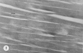
图1 死后8h大鼠肌肉,肌纤维呈节段性膨大,尤如“爆裂的香肠”。PTAH染色,×200
, 百拇医药
Figure 1 Sample of rat muscle in postmortem 8 hrs. The segmental swollen of muscle fibers were seen.PTAH staining, ×200
图2 死后16h大鼠肌肉,肌纤维横纹清晰。PTAH染色,×400
Figure 2 Sample of rat muscle in postmortem 16 hrs. The structure of muscle striations was keep well.PTAH staining, ×400
(二)酶组织化学染色
, http://www.100md.com
SDH:死后立即取材的肌肉,横切肌纤维见肌质网平行排列,整齐,Ⅰ型纤维染色深,Ⅱ型浅淡;死后2~4h,其染色程度变化不大(图3);6h可见肌质网上有间隔的横纹,Ⅱ型纤维呈颗粒状;死后12h肌质网融合,紊乱;16小时肌丝松散,呈波浪状(图4);死后24h与16h变化相似。
图3 死后4h大鼠肌肉SDH染色,Ⅰ型纤维染色深,肌浆网呈平行排列,Ⅱ型纤维染色浅。×400
Figure 3 Sample of rat muscle in postmortem 4 hrs.The I Subtype muscle fibers were obviously stained with parallel of cytoreticulum,the Ⅱ subtype muscle fibers were slightly stained.SDH staining,×400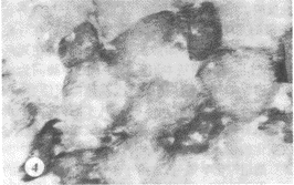
, http://www.100md.com
图4 死后16h大鼠肌肉SDH染色,Ⅰ型纤维染色有所变浅,但不显 著,肌丝呈波浪状。×400
Figure 4 Sample of rat muscle in postmortem 16 hrs. The tissue staining of Ⅰ subtype muscle fibers were gradually decreased. The changes of wave myofibrile were seen.PTAH staining, ×400
NADHD:染色程度变化与SDH相似,但酶染色主要出现在肌细胞膜边缘。死后16和24h,均可 见肌丝松散,染色淡,可见横纹(图5)。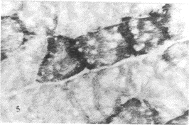
, http://www.100md.com
图5 死后16h大鼠肌肉NADH染色,Ⅰ型肌纤维染色变浅,阳性染色 主要位于肌膜边缘。×400
Figure 5 Sample of rat muscle in postmortem 16 hrs. The tissue staining of Ⅰ subtype muscle fibers were obviously decreased, but the sarcolemma was positive in staining.NADH staining, ×400
CCO:Ⅰ型纤维染色浅淡,Ⅱ型纤维较强。死后8h,Ⅰ型纤维染色即呈阴性(图6);24h则仅 余下Ⅱ型纤维周边阳性染色。
, http://www.100md.com 图6 死后8h大鼠肌肉CCO染色,Ⅱ型纤维染色均较浅,I型纤维染 色几乎完全消失。×400
Figure 6 Sample of rat muscle in postmortem 8 hrs. The tissue staining of Ⅱ subtype muscle fibers were slight,Ⅰ subtype muscle fibers were almost disappeared. CCO staining, ×400
讨 论
在法医学实践中,常根据死后改变推测死亡时间,常用的是早期尸体现象,尸僵是其中之一。对尸僵的肉眼观察报告较多,但对形成尸僵的基本单位--肌纤维的研究则并不多见。
骨骼肌的酶相当丰富,其含量最多的是ATP酶,氧化还原酶类和磷化酶。细胞色素氧化酶和脱氢酶同属于氧化还原酶类,根据其作用底物的化学基团或化学键的不同而分为若干亚类。根据不同肌纤维酶的活性不同将肌纤维分为Ⅰ型和Ⅱ型两大类。
, 百拇医药
本研究通过对大鼠肌肉死后不同时间酶的变化的观察,发现死后SD H和NADHD变化幅度较小,均为Ⅰ型纤维染色强,Ⅱ型纤维染色浅淡,常在死后24h酶的活性仍存在,Ⅱ型纤维均呈阳 性反应。细胞色素氧化酶(CCO)在肌纤维的分布与SDH和NADHD相似,不同的是CCO在骨骼肌的活性程度远不如前两种酶强,因此,在死后16小时Ⅱ型纤维基本均呈阴性。三种酶同为线粒体内膜的标志酶,并且与线粒体内膜结合,只有细胞发生自溶,线粒体破坏时才消失,但由于其在肌纤维上分布量的差异,即CCO含量较小,故变化较快。这与过去学者观察的结果相似[3]。
酶组织化学染色,常由于条件不一致,染色的强度则发生差异,本研究为了避免该 方法的这一缺点,采用不同时间组的动物组织包埋在一起的方法,使其切片厚度,孵育液PH值和孵育时间长短均处于同一条件下,使最后在比较染色结果时排除人为的干扰。因此,结果较可靠。由于日前所用的酶组织化学染色在观察结果时常采用半定量方法,有一定缺陷,如能与图像分析相结合,在判断死亡时间则更具有价值,这将在今后进一步研究。
, http://www.100md.com
*四川省公安厅科研基金资助项目
参考文献
1.Liao ZG,et al. Estimation of postmortem interval based on the changes of phosphorylase activity in skeletal muscles. The Proceedings of XVI international Conference of Forensic Medicine and Legal medicine, Strasbourg, France, 1994:156
2.廖志钢,易旭夫,张永亮,等.死后不同时间大鼠肌肉的扫描电镜观察.华西医科大学报,1998,29(3):323
3.Janssen W. Forensic pathology. Sprinser-Verlag. Berlin,Heidelkerg,New York,Tokyo 1984:38
(收稿:1999-04-05,修回:1999-06-30), 百拇医药