视网膜色素上皮―脉络膜在近视发病中的作用
作者:胡诞宁 Steven A.McCormick
单位:美国纽约医学院 纽约眼耳鼻喉科医院
关键词:近视;病理生理学;生长因子;病理生理学;视网膜色素上皮细胞;病理生理学;黑素细胞;病理生理学;成纤维细胞;病理生理学
眼视光学杂志000401 [摘 要] 目的:探讨人眼视网膜色素上皮细胞(RPE)、黑素细胞、成纤维细胞与碱性成纤维细胞生长因子(bFGF)、β转化生长因子(TGF-β)和肝细胞生长因子(HGF)的关系。方法:①用不含血清的培养液培养三种细胞48h,收集培养液,用酶标免疫吸附法测量生长因子含量;②将各种生长因子加入含有三种不同细胞的培养液中,五天后行细胞计数。 结果:在RPE和成纤维细胞培养液中能测出HGF、bFGF和TGF,而在黑素细胞培养液中则不能测出。bFGF能刺激上述三种细胞的生长,HGF能刺激RPE与黑素细胞的生长,而TGF-β能抑制三种细胞的生长。结论:RPE、成纤维细胞和黑素细胞具有上述生长因子受体,其中RPE和成纤维细胞还能产生上述生长因子。视网膜产生的各种与近视有关的信使不能透过RPE―脉络膜,所以不能直接作用于巩膜。RPE、成纤维细胞和黑素细胞通过产生、抑制和应答各种生长因子、神经递质及激素,在近视发生中产生作用。
, http://www.100md.com
[中图分类号] R778.1+1 [文献标识码] A
[文章编号] 1008-1801(2000)04-0197-04
Role of RPE-Choroid axis on the occurrence of myopia
HU Dan-ning,Steven A. McCormick.
(Tissue Culture Center, New York Eye and Ear Infirmary, New York Medical College)
Abstract: Objective:To study the production of and response to three growth factors related to the occurrence of myopia basic fibroblast growth factor (bFGF), transforming growth factor-β(TGF-β) and hepatocyte growth factor (HGF) by the cell components of RPE-choroid axis (RPE, melanocytes and fibroblasts) in vitro.Methods:RPE, uveal melanocytes and choroidal fibroblasts were isolated and cultured as previously reported. ①Early passages of cells were cultured with serum-free medium for 48 hours. The conditioned media were collected and the amounts of various growth factors were measured by immunoassay. ②Various growth factors were added to the culture media of these cells. After 5 days, cells were counted and compared to the controls.Results:HGF(37-625pg/ml),bFGF(5-34pg/ml) and TGF-β(359-3271pg/ml) were detected in the conditioned media of RPE and fibroblasts, but not in that of melanocytes. bFGF stimulated the growth of RPE, fibroblasts and melanocytes. HGF stimulated the growth of RPE and melanocytes. TGF-β inhibited the growth of all these cells.Conclusion:RPE, fibroblasts and melanocytes have the receptors of these growth factors. RPE and fibroblasts also can produce these growth factors. The present and previous studies indicate that various retina-originated messengers related to the occurrence of myopia cannot pass through the RPE-choroid without interruption to influence the sclera directly. RPE, fibroblasts and melanocytes play an important role in the occurrence of myopia via the production, binding and response to various growth factors, neurotransmitters and hormones.
, http://www.100md.com
Key words: myopia/physiopathology; growth factor/physiopathology; retinal pigment epithelium(RPE)/physiopathology; melanocyte/physiopathology; fibroblast/physiopathology
视网膜色素上皮(retinal pigment epithelium,RPE)―脉络膜在近视发病中所起的作用,历来很少有人研究[1]。实验动物近视研究中发现诱使近视发生的物质主要来自神经视网膜。在近视形成过程中,各种视网膜产生的生化物质(如神经递质、激素、生长因子等)的制造会有所增加或减少[2~4]。过去一般认为此类生化物质(可称为近视信使)能畅行无阻地通过RPE和脉络膜到达巩膜,从而促进或抑止近视之生成。但我们的研究发现脉络膜黑素细胞(melanocyte)具有多种近视信使之受体,从而可阻断其作用[5,6]。因此我们(1998)提出一假设,即近视信使从视网膜产生后先作用于黑素细胞,产生新的信使再作用于巩膜,影响近视之发生[1]。
, http://www.100md.com
在已知的近视信使中有两种生长因子可影响实验近视之发生,即碱性成纤维细胞生长因子(basic fibroblast growth factor,bFGF)与β转化生长因子(transforming growth factor-β,TGF-β)[2,3,7]。在分子生物学研究中又发现肝细胞生长因子(hepatocyte growth factor, HGF)能影响眼球之生长[8]。
我们已建立了人眼RPE、脉络膜黑素细胞与成纤维细胞的分离培养方法及体外试验模型[9~13]。本研究旨在探讨:①体外培养中以上三种细胞制造bFGF、HGF、TGF-β之能力。②该三种生长因子对上述三种细胞之作用。
1 对象和方法
人眼RPE细胞按我们已报告之方法进行分离培养,用加有10%胎牛血清及加2mM谷氨酰胺(gltamine)的F12培养液培养[9]。人眼脉络膜黑素细胞用我们报告的酶梯次分离法进行分离,用加有10%胎牛血清、50ng/ml的12-0-tetradecancyl-phorbol-13-acetate(TPA)、0.1mM的isobutylmethylxanthine与10ng/ml cholera toxin的F12培养液培养[10]。人眼成纤维细胞系在去除贴附的RPE后用酶分离法从脉络膜分离,用含10%胎牛血清的DMEM培养液培养。细胞鉴定系用免疫细胞化学方法,RPE能表达S-100与细胞角蛋白(cytokeratin),黑素细胞仅表达S-100,成纤维细胞则两者均不表达。
, http://www.100md.com
测试生长因子的制造时,用不含血清的培养液培养三种细胞48小时,收集培养液,用酶标免疫吸附测定法(ELISA)测量bFGF、TGF-β2与HGF之含量。测试生长因子作用时,将三种细胞接种到24皿板,每皿接种1×10⁴细胞。次日将三种生长因子分别加入。测试浓度为:bFGF,30ng/ml;HGF,100ng/ml;TGF-β2,3ng/ml。以不加生长因子之培养作对照。培养5天后将细胞脱下,计数并与对照组比较,以t-test测定其差别之显著性。
2 结果
RPE培养液、成纤维细胞培养液、黑素细胞培养中三种生长因子的浓度见表1。
表1 三种培养液中三种生长因子的浓度 (pg/ml)
Tab.1 The concentration of three types of
, 百拇医药
growth factors in the three types of culture
PPE culture
fibroblast culture
melanocyte culture
bFGF
10~15
5~34
-
HGF
37~112
537~625
, 百拇医药
-
TGF-β2
1902~3271
359~579
-
bFGF能刺激RPE、黑素细胞及成纤维细胞之生长,HGF能刺激RPE与黑素细胞之生长,TGF-β2能抑制该三种细胞之生长(图1)。
图1 各种生长因子对培养的色素上皮细胞、脉络膜黑素细胞
及成纤维细胞的影响。与对照组比较,**P<0.01
Fig.1 Effects of various growth factors on the growth of RPE,uveal melanocytes and fibroblasts in vitro.
, 百拇医药
3 讨论
近年来近视之药物治疗徘徊不前,仅有的有效药物阿托品[覃毒硷(muscarine)对抗药]也是在临床研究中发现能防止近视之进行。动物实验中发现了很多种视网膜制造的生长因子(bFGF、HGF、TGF-β)、激素[褪黑激素(melatonin)、5-羟色胺(serotonin)、vasoactive intestine polypeptide(VIP)]和神经递质[覃毒硷、多巴胺(dopamine)]均能促进或抑止近视之发生[2~4]。按理说这些发现必将促进近视的药物治疗发展。但迄今为止,此类物质或其对抗药物均未能用于近视之药物治疗,原因何在值得探讨。
我们发现脉络膜黑素细胞具有褪黑激素与覃毒硷等近视信使之受体[5,6],因此提出假设:由于黑素细胞具有近视信使之受体,因此此类物质无法直接到达并作用于巩膜,对近视发挥作用[1]。
, http://www.100md.com
实验研究发现,近视发生时,眼部bFGF含量下降而TGF-β2含量上升。眼部注射bFGF能抑止近视之发生,而TGF-β2能对抗bFGF,有防止近视作用[2,7]。在鼠眼球生长基因研究中,发现制造HGF之基因可能与眼球生长密切相关,因此HGF也可能影响近视之发生[8]。
生长因子在眼部之产生部位尚无定论,一般认为可能在视网膜产生[14~15]。本项研究发现RPE与脉络膜成纤维细胞均能制造bFGF、HGF、与TGF-β2,提示影响近视发生之生长因子也可能源自RPE与脉络膜。bFGF、HGF与TGF-β2能影响RPE、黑素细胞与成纤维细胞之生长,表明这些细胞也必有此类生长因子的受体存在。
表2 近视信使的受体在色素上皮―脉络膜细胞中存在的情况
, 百拇医药 Tab.2 The presence of receptors of retina-originated messengers related to
myopia in the cells located in the RPE-choroid
RPE
Melanocytes
Fibroblasts
Dopamine(Ref.26)
+
?
?
Melatonin(Ref.11,27)
, http://www.100md.com
+
+
?
Serotonin(Ref.28)
+
?
?
bFGF
+
+
+
HGF
+
, 百拇医药 +
-
TGFβ
+
+
+
Muscarine(Ref.10)
?
+
?
VIP(Ref.29)
+
?
, 百拇医药
?
Nitric Oxide(Ref.1)*
+
+
?
*It has been demonstrated that melanin in the cultured RPE and melanocytes can reduce the amount of exogenous nitric oxide in the culture medium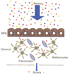
图2 视网膜产生的信使阻止位于色素上皮一脉络膜的各类细胞,使它们不能到达巩膜,从而阻滞或减轻近视发生的作用。
, http://www.100md.com
Fig.2 Retina-originated messengers bind to various cell types located in the
RPE-choroid,so that they cannot enter the sclera to express they effects.
Their effects are blocked or reduced.
图3 视网膜产生的信使激发色素上皮-脉络膜上的各类细胞释
放其他信使作用于巩膜而影响近视的发生。
Fig.3 Retina-oringinated messengers stimulate various cell types located in the
, http://www.100md.com
RPE-choroid to release other messengers to affect the sclera
and play a role in the occurrence of myopia.
进一步复习文献及回顾我们过去的研究,发现RPE、黑素细胞与成纤维细胞具有上述所有能影响近视的生长因子、激素及神经递质的受体(表2)[1,5,6,19~20]。因此证明并扩大了我们假设的范围,即已知的由神经视网膜制造的与近视有关的生化物质,无一能顺利透过RPE与脉络膜而直接作用于巩膜。RPE与脉络膜内各种细胞之作用可能为:①这些细胞可与各种近视信使结合,阻断其作用(图2)。②各种近视信使与RPE等细胞结合后能刺激或抑止这些细胞制造与近视有关的生化物质,从而作用于巩膜而影响近视之发生(图3)。RPE与脉络膜对近视的确切作用还有待研究。如能了解经RPE和脉络膜级联系统(cascade)产生的最终近视信使,必能为近视的药物治疗提供重要信息。因此,对RPE―脉络膜与近视关系的深入研究,除有助于了解近视之发病机制外,并可为近视药物治疗带来新的希望。
, 百拇医药
基金项目:Supported by the New York Eye and Ear Infirmary Departments of Ophthalmology and Pathology Research Funds。
作者简介:胡诞宁,男,上海人,旅美学者,纽约医学院眼科教授、纽约眼耳鼻喉科医院组织培养研究中心主任。
参考文献
[1] Hu DN,Roberts JE,McCormick SA. Role of uveal melanocytes in the development of myopia[A]. In Myopia Updates Ⅱ(Proceedings of the Ⅶ International Conference on Myopia)[M]. Tokyo:Springer,2000.125-126.
, 百拇医药
[2] Seko Y,Shimokawa H,Tokoro T. Expression of bFGF and TGF-β2 in experimental myopia in chicks[J]. Invest Ophthalmol Vis Sci,1995,36:1183-1187.
[3] Honda S,Fujii S,Sekiya Y,et al. Retinal control on the axial length mediated by transforming growth factor-β in chick eye[J]. Invest Ophthalmol Vis Sci,1996,37:2519-2526.
[4] Bitzer M,Schwan H,Schaeffel F. Effects of atropine on ZENK expression in the chicken retina. In Myopia 2000(Proceedings of the Ⅷ International Conference on Myopia)[M]. Boston:Conference on Myopia 2000,Inc. 2000,211-212.
, http://www.100md.com
[5] Hu DN,Woodward DF,McCormick ST. Influence of autonomic neurotransmitters on human uveal melanocytes in vitro[J]. Exp Eye Res,2000,71:217-224.
[6] Roberts JE,Wiechmann AF,Hu DN. Melatonin receptors in human uveal melanocytes and melanoma cells[J]. J Pineal Res,2000,28:165-171.
[7] Rohrer B,Stell WK. Basic fibroblast growth factor(bFGF) and transforming growth factor beta(TGF-β) act as stop and go signals to modulate postnatal ocular growth in the chick[J]. Exp Eye Res,1994,58:553-562.
, 百拇医药
[8] Zhou G,Williams RW. Eye1 and Eye2:Gene loci that modulate eye size,lens weight,and retinal area in the mouse[J]. Invest Ophthalmol Vis Sci,1999,40:817-825.
[9] Hu DN,Del Monte D,Liu S,et al. Morphology,phagocytosis,and vitamin Ametabolism of cultured human retinal pigment epithelium[J]. Birth Defect,1982,18(6):61-79.
[10] Hu DN,McCormick SA,Ritch R,et al. Studies of human uveal mel-
anoctyes in vitro:Isolation,purification and cultivation of human uveal melanocytes[J]. Invest Opththlmol Vis Sci,1993,33:2210-2219.
, 百拇医药
[11] Hu DN,McCormick SA,Ritch R. Studies of human uveal melanocytes in vitro:Growth regulation of cultured human uveal melanocytes[J]. Invest Ophthalmol Vis Sci,1993,34:2220-2227.
[12] Hu DN,McCormick SA,Orlow SJ,et al. Regulation of melanogenesis by human uveal melanocytes in vitro[J]. Exp Eye Res,1997,64:397-404.
[13] Hu DN,Stiernschantz J,McCormick SA. Effect of prostaglanding A2,E1,F1α and latanoprost on cultured human iridal melanocytes[J]. Exp Eye Res,2000,70:113-120.
, http://www.100md.com
[14] Wallman J. Retinal influence on scleral underlie visual deprivation myopia. In:Bock G,widdows K,eds. Myopia and the Control of Eye Growth. Ciba Foundation Symposium 155[M]. Chichester:John Wiley and Sons,1990.126-135.
[15] Ikeda T,Nishimura M,Ushiyama M,et al. Vitreous levels of human hepatocyte growth factor increase in proliferative diabetic retinopathy[J]. Invest Ophthalmol Vis Sci,1998,39(4):S124.
[16] Hackett SF,Schoenfeld CL,Freund J,et al. Neurotrophic factors,cytokines and stress increase expression of basic fibroblast growth factor in retinal pigment epithelial cells[J]. Exp Eye Res,1997,64:865-873.
, http://www.100md.com
[17] Mertz JR,Wallman J,Choroidal retinoic acid synthesis:A possible mediator between refractive error and compensatory eye growth[J]. Exp Eye Res,2000,70:519-527.
[18] Guggenheim J,McBrien NA. Form-deprivation myopia induces activation of scleral matrix metalloproteinase-2 in tree shrew[J]. Invest Ophthalmol Vis Sci,1996,37:1380-1395.
[19] Haque R,Maltseva O,Ivanova T,et al. Dopamine D1 recpptor expression in cultured human and monkey retinal pigment epithelial cells[J]. Invest Ophthalmol Vis Sci,2000,41:S843.
[20] Nash M,Flanigan T,Leslie R,et al. Serotnin-2A receptor mRNA expression in rat retinal pigment epithelial cells[J]. Ophthalmic Res,1999,31:1-14.
收稿日期:2000-08-09, 百拇医药