作者:徐洪涛 姜忠良 朴春爱 王文兴
单位:徐洪涛 姜忠良(中国科学院海洋研究所,山东 青岛 266071);朴春爱(TAMU,TX77843,USA);王文兴(国家海洋局第一海洋研究所,山东 青岛 266003)
关键词:牙鲆;虹彩病毒;淋巴囊肿病;DNA序列测定
病毒学报000307 摘要:近几年我国海水养殖鱼类发生病毒性传染病,病鱼鳍、鳃及体表皮肤出现乳头瘤样赘生物。将发病牙鲆(Paralichthys olivaceus)表皮瘤组织进行超薄切片,电镜下在病变组织明显肥大的细胞胞浆内发现大量球状病毒,呈二十面体对称,具包膜,直径约210nm。根据虹彩病毒主要衣壳蛋白(major capsid protein,MCP)基因中间保守区序列设计简并引物,应用PCR方法从病变组织中扩增出长636bp的片段。经DNA序列测定及同源性分析表明,该段核苷酸序列与欧洲分离的淋巴囊肿病病毒1型(lymphocystic disease virus type 1,LCDV-1)MCP基因相应序列有77%的同源性,表明该病毒属虹彩病毒科(Iridoviridae)淋巴囊肿病毒属(Lymphocystivirus),但与欧洲分离株有一定差异。将该病毒暂命名为淋巴囊肿病病毒中国分离株(lymphocystis disease virus-Chinese isolate,LCDV-cn)。
中图分类号:S852.65
+9.1;Q523
+.8 文献标识码:A
文章编号:1000-8721(2000)03-0223-04
Study on the Causative Agent of Lymphocystic Disease in Cultured Flounder, Paralichthys Olivaceus, in Mainland China
XU Hong-tao,JIANG Zhong-liang
(Institute of Oceanology, Chinese Academy of Sciences, Shandong, Qingdao 266071, China)
PIAO Chun-ai
(TAMU, TX 77843, USA)
WANG Wen-xing
(First Institute of Oceanography, National Bureau of Ocean, Shandong, Qingdao 266003, China)
Abstract: Viral epizootic causing papilloma-like lesions on the skin, gill and fin of factory-cultivated flatfish, Paralichthys olivaceus, occurred in mainland China and caused great economic loss in the past few years. Electron microscopy showed large amount of enveloped icosehedral, hexogonal viruses in the cytoplasm of hypertrophied cells. The diameter of the virus averages in 210 nm (side to side) . A pair of degenerate primers based on the conserved sequence of the major capsid protein (MCP) gene of iridovirus amplified a 636bp fragment using DNA extracted from the virus purified from diseased fish tissues as the template. The nucleotide sequence of the amplified fragment showed 77% homology with the corresponding region of the MCP gene of lymphocystic disease virus type 1 (LCDV-1) . Both elcectron microscopy and DNA sequencing results showed that this virus belongs to genus Lymphocystivirus, family Iridoviridae. The virus reported here was named temporally the Chinese flounder isolate of lymphocystic disease virus (LCDV-cn) . This is the first report on the partial DNA sequencing of the MCP gene of piscine iridovirus isolated in mainland China.
Key words: Paralichthys olivaceus; Iridovirus; lymphocystic disease; DNA sequencing
海水养殖业一直是我国国民经济的支柱产业之一。近几年养殖的牙鲆(Paralichthys olivaceus)、鲈鱼、真鲷、石斑鱼等名贵鱼种发生一种病毒性传染病,病鱼体表呈现乳头瘤样赘生物,感染率在80%以上,死亡率近30%
[1,2],严重影响其市场价值,造成重大经济损失。牙鲆是我国工厂化养殖的主要鱼类品种,为查明该病的病原特征,我们对发病牙鲆进行了电镜观察,在病变组织细胞浆内发现虹彩病毒样颗粒。根据虹彩病毒主要衣壳蛋白(major capsid protein,MCP)基因中间保守区序列设计引物,从病变组织中扩增出长为636bp的DNA片段,并进行了序列测定。
材料与方法
1 标本 患病牙鲆于1997年9月10日取自山东荣成邱家养殖场,由国家海洋局第一海洋研究所王文兴教授提供,冻存于-70℃。健康牙鲆为国家海洋局第一海洋研究所存养。
2 电镜标本制备 取自然发病牙鲆的表皮瘤组织经2.5%戊二醛固定、1%锇酸后固定、乙醇脱水、Epon812树脂包埋后,进行常规超薄切片,醋酸铀染色,透射电镜观察、拍照。
3 病毒分离纯化及基因组DNA提取 病毒纯化参照文献
[3-6]的方法并加以改进:取5g表皮瘤组织,于50ml TNE
[7]缓冲液中匀浆,3?000r/min离心20min,上清铺于30%蔗糖垫层,25?000r/min(Beckman SW27转子)。4℃离心120min,沉淀用适量TNE缓冲液重悬,上10%~35%氯化铯密度梯度,30?000r/min(Beckman SW40转子)10℃离心24h,取乳白色病毒带对TNE缓冲液4℃透析36h。纯化病毒核衣壳悬液加蛋白酶K及Sarkosyl分别至0.1mg/ml及0.5%,56℃孵育2h,酚、酚:氯仿及氯仿各抽提1次,乙醇沉淀,溶于TE用作PCR反应模板。
4 健康牙鲆表皮组织DNA提取 按文献
[7]所述真核细胞基因组DNA提取方法进行,用作PCR反应阴性对照模板。
5 PCR扩增反应 简并引物系依据虹彩病毒MCP中间保守区序列
[8-11]设计,由中国科学院微生物研究所合成:
正向引物:5′ATGAT(T/C)GG(T/A)AAT(A/G)C(A/T)(A/T)(T/C)T3′
反向引物:5′CA(C/G)CAAA(A/G)AATAATA(T/A)TCA(C/G)(T/A)AC3′
50μl PCR反应混合物内含:引物各50pmol,dNTPs各250μmol/L,MgCl
2 1.5mmol/L,Taq DNA聚合酶2U,DNA模板适量。反应在PE-2400热循环仪上进行,循环参数为:94℃变性5min;94℃45s,45℃45s,72℃45s,循环30次;72℃延伸7min。
6 PCR产物克隆及DNA序列测定 PCR产物经纯化后连接到pGEM-T载体(Promega产品),转化致敏菌DH5α,提取质粒DNA。序列测定由宝生物(大连)生物工程有限公司完成。
7 DNA序列同源性分析 应用Dnasis序列分析软件进行。
结 果
1 发病牙鲆病变特征
在发病牙鲆体表皮肤、鳃、鳍等部位出现灰白色乳头瘤样赘生物,部分肿瘤组织聚集成桑椹状(图1)。严重者赘生物遍布全身,甚至挤出病鱼眼球。病变也可见于咽部、肠系膜、肝、脾和卵巢。

图1 患病牙鲆表皮瘤
Figure 1 Epidermic tumor of diseased Paralichthys olivaceus
2 电子显微镜观察
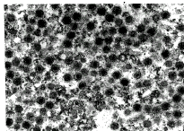
病变组织细胞浆内可见大量六角形立体对称病毒颗粒,有包膜,完整毒粒直径约为210nm(边-边),大量毒粒堆积可呈晶格状排列(图2)。

图2 患病牙鲆病变组织超薄切片的电镜照片(×41?340)
Figure 2 Electron micrograph of the tumor tissue from diseased Paralichthys olivaceus(×41,340)
3 PCR扩增产物序列测定
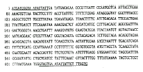
以纯化病毒DNA为模板扩增出长约0.6kb的DNA片段,扩增片段经纯化、克隆和序列测定获得长为636bp的核苷酸序列(图3)。健康牙鲆组织DNA无相应扩增产物。
4 核苷酸序列同源性分析
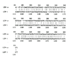
本文报道的病毒(以LCDV-cn表示)基因序列与淋巴囊肿病病毒1型(LCDV-1)MCP基因
[9]631~1?266位核苷酸同源性为77%(图4)。

图3 PCR扩增产物DNA序列
划线部分示引物区
Figure3 DNA sequence of the PCR amplification product
The primer regions are underlined.


图4 LCDV-cn及LCDV-1 MCP基因部分核苷酸序列同源性分析
Figure 4 Nucleotide homology analysis of partial DNA sequence of MCP gene of LCDV-cn and LCDV-1
讨 论
虹彩病毒科(Iridoviridae)是一组从多种昆虫及脊椎动物宿主中分离到的二十面体对称的胞浆内DNA病毒,分为五个病毒属
[12],即:虹彩病毒属(Iridovirus)、绿虹彩病毒属(Chloriridovirus)、蛙病毒属(Ranavirus)、淋巴囊肿病毒属(Lymphocystivirus)和金鱼病毒1型样病毒属(Goldfish virus 1-like viruses)。该科病毒具有独特的形态学特征,病毒呈六角形、二十面体对称,有包膜,在细胞浆内装配。根据本文报道的发病牙鲆病变组织超薄切片的电子显微镜观察结果,该病毒符合上述典型的形态学特征,属虹彩病毒科。尽管仅仅根据形态学特征很容易确定一种病毒是否为虹彩病毒,但不能确定病毒属于何种病毒属,也不能确定从不同宿主或不同地域内的同类宿主中分离的病毒是否为同一种病毒或变异株,本文依据MCP是虹彩病毒的主要结构蛋白,在胞浆内双链DNA病毒,如虹彩病毒、藻类DNA病毒及非洲猪瘟病毒中具有高度保守性,是研究病毒进化及分类的良好对象
[11],目前广泛应用于新分离的脊椎动物虹彩病毒的分类鉴定。通过MCP基因序列同源性分析,最近已发现新分离的引起鱼类系统性感染的虹彩病毒属于蛙病毒属。我们根据虹彩病毒MCP基因中间高度保守区设计引物,从国内养殖的发病牙鲆病变组织中扩增出长为636bp的片段,经序列测定及同源性分析表明,其核苷酸序列与欧洲分离的淋巴囊肿病病毒1型(LCDV-1)MCP基因631~1266位核苷酸序列同源性为77%,表明扩增产物确为病毒的基因序列而非非特异性扩增产物。LCDV-1是淋巴囊肿病病毒属的代表株,因此可以确定本文研究的病毒属于淋巴囊肿病病毒属,但与LCDV-1存在差异,是不同的分离株,提示相同病毒在不同地域内的相似宿主中存在变异株。为别于欧洲分离的LCDV-1,暂将其命名为淋巴囊肿病病毒中国分离株(lymphocystic disease virus-Chinese isolate,LCDV-cn)。
本文对我国养殖牙鲆淋巴囊肿病病毒进行了电子显微镜观察,并测定了其MCP中间保守区636bp的核苷酸序列,不仅明确了该病毒的基本特征,同时表明LCDV-cn与欧洲分离的LCDV-1存在一定差异,说明从不同地域中同类宿主中分离的病毒毒株不同。根据该段DNA序列设计特异性引物,建立了LCDV-cn的PCR检测技术(结果另文报道),可以为国内该病毒病的诊断及检疫提供良好的手段。同时,可以用作DNA探针进行LCDV-cn MCP全基因的克隆及鉴定,进而为研制基因工程疫苗打下基础。
作者简介:徐洪涛(1965-),男,医学博士,研究海洋病毒学。
本文报道的序列已由GenBank发布,录入号BankIt 252036。
参考文献:
[1]周丽,宫庆礼,编著.海水鱼虾蟹贝病害防治技术[M].青岛:青岛海洋大学出版社,1998.
[2]孟庄显,俞开康,编著.鱼虾蟹贝疾病诊断和防治[M].北京:中国农业出版社,1996.
[3] Darai G, Anders K, Koch H G, et al. Analysis of the genome of fish lymphocystic disease virus isolated directly from epidermal tumours of pleuronectes [J] . Virology, 1983, 126 (2) : 466-479.
[4] Samalecos C P. Analysis of the structure of fish lymphocystic disease virions from skin tumours of pleuronectes [J] . Arch Virol, 1986, 91 (1-2) : 1-10.
[5] Robin J, Berthiaume L. Purification of lymphocystic disease virus (LCDV) grown in tissue culture. Evidences for the presence of two types of viral particles [J] . Rev Can Biol, 1981, 40 (4) : 323-329.
[6] Berthiaume L, Alain R, Robin J. Morphology and ultrastructure of lymphocystisc disease virus, a fish iridovirus, grown in tissue culture [J] . Virology, 1984, 135 (1) : 10-19.
[7] Sambrook J, Fritsch E F, Maniatis T. Molecular Cloning: A Laboratory Manual [M] . 2nd ed. New York, USA: Cold Spring Harbor Laboratory Press, 1989.
[8] Mao J, Tham T N, Gentry G A, et al. Cloning, sequence analysis and expression of the major capsid protein of the iridovirus frog virus 3 [J] . Virology, 1996, 216 (2) : 431-436.
[9] Schnitzler P, Darai G. Identification of the gene encoding the major capsid protein of fish lymphocystic disease virus [J] . J Gen Virol, 1993, 74 (10) : 2143-2150.
[10] Stohwasser R, Raab K, Schnitzler P, et al. Identification of the gene encoding the major capsid protein of insect iridescent virus type 6 by polymerase chain reaction [J] . J Gen Virol, 1993, 74 (5) : 873-879.
[11] Tidona C A, Schnitzler P, Kehm R, et al. Is the major capsid protein of iridoviruses a suitable target for the study of viral evolution [J] ? Virus Genes, 1998, 16 (1) : 59-66.
[12] Murphy F A, Fanquet C M, Bishop D H L, et al. Virus taxonomy, 6th report of the International Committee on Taxonomy of Viruses [C]. Arch Virol, 1995, Suppl. 10: 208-239.
[13] Mao J, Hedrick R P. Chinchar V G. Molecular characterization, sequence analysis, and taxonomic position of newly isolated fish iridoviruses [C] . Virology, 1997, 229 (1) : 212-220.
收稿日期:1999-03-03;修回日期:1999-05-11
婵犵數濮烽弫鍛婃叏閻戣棄鏋侀柛娑橈攻閸欏繘鏌i幋锝嗩棄闁哄绶氶弻鐔兼⒒鐎靛壊妲紒鐐劤濞硷繝寮婚悢琛″亾閻㈢櫥鐟扮摥缂傚倷娴囩紙浼村磹濠靛钃熼柨鏇楀亾閾伙絽銆掑鐓庣仭闁挎稑妫楅埞鎴︽倷閸欏鏋欐繛瀛樼矋缁诲牆鐣烽幋锕€绠涢柣妤€鐗嗘禒顖炴⒑閹肩偛鍔村ù婊勭矒閹啴鎮滃Ο闀愮盎闂佹寧妫侀褔鐛弽顓熺厓閻熸瑥瀚悘鎾煙椤旇娅囩紒杈ㄥ笒铻e┑鐘崇閺嗘ê鈹戦悩鍨毄闁稿鐩幆鍥ㄥ閺夋垹锛欏┑鐘绘涧椤戝啴鍩€椤戣法顦︽い顐g箞閹虫粓鎮介棃娑樞﹂梺璇查缁犲秹宕曢崡鐐嶆稑鈽夐姀鐘插亶闂佸憡顨堥崑鎰板绩娴犲鐓熼柟閭﹀幗缂嶆垿鏌h箛瀣姢闁逞屽墲椤煤閿曞倸绀堥柣鏂款殠閸ゆ洟鏌熼梻瀵割槮閸ユ挳姊虹化鏇炲⒉闁告梹甯為懞杈ㄧ節濮橆厸鎷洪梺鍛婂姇瀵爼骞嗛崼銉︾厱闁绘洑绀佹禍鎵偓瑙勬磸閸ㄤ粙鐛鈧、娆撴偩鐏炶棄绠伴梻浣筋嚙缁绘帡宕戝☉娆愭珷濞寸姴顑呴悡鈥愁熆閼搁潧濮堥柣鎾冲暣閺岋箑螣娓氼垱鈻撳┑鈽嗗灙閸嬫捇鏌f惔銈庢綈婵炴祴鏅濋幑銏犫攽鐎n亞鐣哄┑鐐叉濞存岸宕崨顔轰簻闁哄倸澧介惌娆撴煟濞戞﹢鍙勬慨濠傤煼瀹曟帒鈻庨幋顓熜滈梻浣告贡閳峰牓宕戞繝鍛棨婵$偑鍊栭崝褏绮婚幋锔藉仾闁绘劦鍓涚弧鈧繝鐢靛Т閸婃悂骞冮埡鍌滅闁瑰瓨鐟ラ悘鈺冪磼閳ь剟宕橀埞澶哥盎闂佸搫鍟ú銈壦夊澶嬬厵闁兼亽鍎茬粈鈧梺瀹狀潐閸ㄥ潡骞冨▎鎾崇骇闁瑰濮冲鎾绘⒑閼瑰府楠忛柛濠冪箞瀵鏁嶉崟銊ヤ壕闁挎繂绨奸柇顖炴煕濞嗗繐顏柡宀嬬秮楠炴瑩宕橀妸銈呮瀳闁诲氦顫夊ú鏍偉閸忛棿绻嗛柟闂寸劍閺呮繈鏌嶈閸撶喖骞嗛崘顔肩闁挎棁袙閹疯櫣绱撴担鍓插剱妞ゆ垶鐟╁畷鏇㈠箛閻楀牏鍘靛┑顔界箓閼活垶宕ラ崷顓犵<妞ゆ梻鈷堥崕鏃€顨ラ悙鏉戞诞鐎殿噮鍓熷畷顐﹀礋椤忓嫷妫滈梻鍌欑劍鐎笛呮崲閸屾粏濮抽柤娴嬫櫈婵啿霉閻樺樊鍎愰柣鎾存礋閺岋繝宕橀埡鍌氭殫闂佷紮缍€妞存悂濡甸崟顖f晜闁告洦鍋呭▓缁樼節绾版ǚ鍋撳畷鍥х厽閻庤娲栧畷顒冪亙闂侀€炲苯澧畝锝呮健楠炲鏁傜憴锝嗗缂傚倷绀侀鍡涱敄濞嗘挸纾块柟鎵閻撴瑩鏌i悢鍝勵暭闁哥姵岣块埀顒侇問閸犳盯顢氳閸┿儲寰勯幇顒夋綂闂佺偨鍎遍崢鏍уΔ鍛厽閹兼番鍊ゅḿ鎰箾閸欏澧悡銈夋煏韫囧鈧洟鎮為崹顐犱簻闁瑰搫绉堕ˇ锔剧磼濡や浇澹橀懣鎰版煕閵夋垵绉烽崥顐︽倵鐟欏嫭绌跨紒鍙夊劤椤曘儵宕熼鍌滅槇闂佺ǹ琚崐鏍礊鐎n喗鈷掗柛灞剧懅椤︼箓鏌i妶鍛枠闁挎繄鍋炲鍕箛椤掆偓閻庮參姊洪崜鑼帥闁哥姵宀稿鎶芥晝閸屾稓鍘卞銈嗗姧缁插墽绮堥埀顒勬⒑缂佹ɑ灏伴柨鏇樺灲瀵鏁愭径濠勭杸闂傚倸鐗婄粙鎴︼綖椤愶絿绠鹃悗娑欋缚閻鏌i悢鍙夋珔妞ゎ偄绻橀幖鍦喆閸曨偆褰撮梻浣告惈鐞氼偊宕曞ú顏勭劦妞ゆ帊绶″▓妯肩磼鏉堛劌娴柟顔规櫊閹崇娀顢栭搴℃噳閸嬫挾鎲撮崟顒傤槰濠电偠灏欓崰搴ㄦ偩閻戣姤鏅搁柣妯哄暱閳ь剟顥撻埀顒€绠嶉崕鍗炍涘☉銏犵叀濠㈣埖鍔栭悡鍐煢濡警妯堟俊顖楀亾濠电姵顔栭崰鎺楀磻閹剧粯鈷戠紓浣癸公閼版寧绻涙担鍐插閸欏繘鏌ㄩ弮鈧崹婵堟崲閸℃稒鐓熼柟鏉垮悁缁ㄥ鏌嶈閸撴岸鎮у⿰鍫濇瀬妞ゆ洍鍋撴鐐村笒椤啫鈹戦崱妯烘灎濡炪們鍨洪惄顖炲箖濞嗘挸绾ч柟瀛樻煥娴滈箖鏌涢锝嗙闁绘挶鍎甸弻锟犲礃閵娿儮鍋撴繝姘闁稿本绋撶粻楣冩煕椤愶絿鐭岄柣顓熺懇閺屸€崇暆鐎n剛袦濡ょ姷鍋涘ú顓€€佸Δ浣瑰闁告瑥顦辨禍鐐烘⒒閸屾瑨鍏岄弸顏堟倵濞戞帗娅呮い顐㈢箺閵囨劙骞掔€Q冧壕闁告劦鍠栧敮闂佸啿鎼崐鎼佸焵椤掑倸鍘撮柡灞剧☉閳诲氦绠涢敐鍠帮箓姊洪崫鍕靛剳濠殿喗鎸抽垾鏃堝礃椤斿槈褔鏌涢幇鈺佸濞寸姵鍎抽埞鎴﹀煡閸℃ぞ绨婚梺鍝ュ櫏閸嬪﹪骞冩ィ鍐╁€婚柦妯猴級閳哄懏鐓冮柛婵嗗閺€濠氭煛閸滃啰绉慨濠呮缁辨帒螣閸濆嫷娼氭繝鐢靛仒閸栫娀宕惰椤旀劗绱撴担鍓插剰缂併劑浜跺銊︾鐎n偆鍘藉┑鈽嗗灥濞咃絾绂掑☉銏$厸闁糕€崇箲濞呭﹪鏌$仦鍓с€掗柍褜鍓ㄧ紞鍡涘礈濞嗘挸鐓濋柛顐ゅ櫏濞堜粙鏌i幇顒傛憼鐎规洖鏈妵鍕閳╁喚妫冮悗瑙勬礀瀹曨剝鐏冮梺鍛婂姦娴滅偤骞楀畝鍕拻闁稿本鐟︾粊鐗堛亜閺囩喓澧电€规洝顫夌缓浠嬪川婵犲倵鍋撻崼鏇熺厵闂侇叏绠戦崝鎺撶箾瀹割喕鎲鹃柡浣稿€块弻娑㈠即閵娿儱瀛i梺鍛婅壘閹虫ê顫忕紒妯诲闁惧繒鎳撶粭锟犳⒑閹稿骸鍝洪柡灞剧☉椤繈鏁愰崨顒€顥氱紓鍌氬€搁崐鐑芥倿閿曞倶鈧啴宕ㄥ銈呮喘濡啫鈽夋潏銊х▉婵犵數鍋涘Ο濠冪濠婂懐涓嶅ù鍏兼綑缁犲綊寮堕崼婵嗏挃闁诡喖銈搁弻锝夋晲閸ャ劍姣堝┑顔硷攻濡炰粙骞冮悜钘夌骇闁圭ǹ宸╅埡渚囨富闁靛牆妫楅悘銉︺亜閿旇鐏﹂柟顕€绠栭幃婊堟嚍閵夈儰缂撶紓鍌欑椤戝懎岣块敓鐘茶埞閻犻缚銆€閺€浠嬫煟濮楀棗鏋涢柣鎺戞惈闇夋繝濠傚缁犳捇鎽堕悙鐑樼厱闁逛即娼ч弸娑㈡煛鐎b晝绐旈柡灞剧〒娴狅箓宕滆閻撳倹淇婇悙棰濇綈濠电偛锕獮鍐ㄎ旈崨顔芥珳闁圭厧鐡ㄧ换鍕儎鎼淬垻绡€缁剧増菤閸嬫捇鎮欓挊澶夊垝婵犳鍠栭敃锔惧垝椤栫偛绠柛娑欐綑瀹告繂鈹戦悩鎻掓殭闁哄鍨垮缁樻媴閸涢潧缍婇、鏍炊椤掆偓绾惧鏌曢崼婵愭Ш鐎规挷绶氶弻鐔兼偋閸喓鍑$紓浣哄閸o綁寮婚敃鈧灒濞撴凹鍨辨瓏闂備胶顢婃慨銈吤洪悢濂夋綎婵炲樊浜滄导鐘绘煕閺囥劌澧柛瀣Ч濮婃椽骞栭悙鎻掝瀳缂傚倸绉撮敃顏堝箖娴兼惌鏁嬮柍褜鍓氶幈銊╁焵椤掑嫭鐓忛柛顐g箖椤ョ娀鏌涘Ο缁樺€愭慨濠冩そ楠炴牠鎮欓幓鎺戭潕闂備礁鎼幊蹇曠矙閹达附鍋ゆい鎾跺亹閺€浠嬫煟濡椿鍟忛柡鍡╁灦閺屽秷顧侀柛鎾寸箘娴滅ǹ鈻庨幘鏉戝壒濠电偛妫欓幐濠氭偂濞戞埃鍋撻崗澶婁壕闁诲函缍嗛崜娑溾叺婵犵數濮甸鏍窗閹烘纾婚柟鍓х帛閳锋垿鎮楅崷顓炐i柕鍡楀暣閺屻倗鈧湱濮撮悡鎰磼閸屾稑娴柛鈺嬬節瀹曟﹢顢旈崱顓犻棷闂傚倷鑳舵灙闁哄牜鍓欓~婵嬪Ω閵壯勬噧闂傚倸鍊烽悞锔锯偓绗涘厾娲晜閻e矈娲稿銈呯箰閻楀﹪宕戝Ο姹囦簻闁哄啫鍊瑰▍鏇犵磼閳锯偓閸嬫捇姊绘担瑙勫仩闁稿寒鍨跺畷婵嗩吋閸涱亝鐎洪梺闈涚箞閸婃牠鍩涢幋鐘电=濞达絽绠嶉埀顒佸浮瀹曠兘顢樺┑鍫濆⒕婵犵數鍋涘Λ娆撳箰婵犳艾纾绘い蹇撶墛閻撳啰鎲稿⿰鍫濈婵﹩鍘鹃埞宥呪攽閻樺弶澶勯柛濠囨敱閵囧嫯绠涢幘鎰佷槐闂佺ǹ顑嗛幑鍥ь嚕娴犲鏁囬柣鎰問閸炶泛鈹戞幊閸娧呭緤娴犲鐤い鎰╁€曢ˉ姘攽閻樺磭顣查柣鎾跺枛閺岋繝宕掑☉姗嗗殝闂佽褰冮幉锟犲Φ閸曨垰鐓涢柛鎰╁妼楠炲姊洪崫鍕伇闁哥姵鐗犻悰顔碱吋婢舵ɑ鏅╃紒鐐妞寸ǹ袙婢舵劖鈷掑ù锝囨嚀閳绘洟鏌¢埀顒勬焼瀹ュ懎鐎梺绉嗗嫷娈旈柦鍐枑缁绘盯骞嬪▎蹇曚痪闂佸搫鎳忕划鎾诲箖瑜版帒鐐婇柕濞垮劤缁侀绱撴担璇℃當闁活厼鍊搁~蹇撁洪鍛闂侀潧鐗嗛幊蹇涙倵妤e啯鈷戦梻鍫氭櫇缁嬬粯绻涢崗鑲╂噮闁告瑥鎳庨—鍐Χ閸涱垳顔囬柣搴㈠焹閸嬫捇鎮峰⿰鍕棃鐎殿喛顕ч埥澶愬閻樻牓鍔嶉妵鍕籍閳ь剟宕曞畷鍥ㄦ殰闁靛鍎弨浠嬫煟閹邦垱纭惧ù婊冪秺閺屾盯鎮╁畷鍥р吂濡炪値鍋勭换鎰弲濡炪倕绻愮€氼噣藝閵娾晜鈷戦柛娑橈功閹冲啯銇勯敂鍨祮鐎规洘娲熼獮妯肩磼濡攱瀚奸梻浣藉吹閸犳劕顭垮鈧铏綇閵婏箑寮垮┑顔筋殔閻楀棝銆呴鍕厵妤犵偛鐏濋悘鈺傘亜椤愶絿绠為柟铏矌閸犲﹤螣鐠囧樊浼滃┑鐘垫暩閸嬬娀骞撻鍡楃筏濞寸姴顑呯粻鐘崇箾閸℃ê绔惧ù婊冪秺閺岀喖鎮滃Ο铏逛淮闂佺粯绻冮敋妞ゎ亜鍟存俊鍫曞幢濡ゅ啰鎳嗛梻浣呵归敃銈夆€﹂悜钘夌畾閻忕偠袙閺嬪酣鏌熼幆褜鍤熼柛妯兼暬濮婅櫣绱掑Ο铏逛桓闂佹寧娲嶉弲娑㈠煝閹捐鍗抽柕蹇ョ磿閸樿鲸绻濋悽闈浶㈤柛濠勭帛缁傛帒鈽夐姀锛勫帾闂佹悶鍎滈崘鍙ョ磾闁诲孩顔栭崳顕€宕滈悢椋庢殾鐟滅増甯╅弫濠囨偡濞嗗繐顏╅柣锕佷含缁辨捇宕掑▎鎺戝帯闂佺ǹ顑嗛幑鍥х暦閺囥垹绠柦妯猴級瑜旈弻鐔兼⒒鐎靛壊妲梺钘夊暟閸犳牠寮婚妸鈺傚亜闁告繂瀚呴姀銏㈢<闁逞屽墴瀹曟﹢顢欓悾灞藉妇濠电姷鏁搁崑娑㈡倶濠靛绀夋俊銈呮噺閻撴洟鎮楅敐搴′簼閻忓繒鏁哥槐鎺楊敊閻e本鍣板Δ鐘靛仦閸旀牠骞嗛弮鍫濐潊闁靛繆妲呭ḿ鐘崇節閻㈤潧啸闁轰礁鎲¢幈銊╂倻閽樺鐎梺鍛婄☉閿曘儵宕h箛鏂讳簻闁规儳宕悘鈺冪磼閻欌偓閸ㄥ爼寮诲☉銏犵疀闂傚牊绋掗悘鍫㈢磽娴f彃浜鹃柣搴秵閸犳鎮¢弴鐔翠簻妞ゆ挾鍠庨悘銉ッ瑰⿰鍛槐闁哄备鍓濆鍕節閸曨厜銊╂⒑閸濆嫮鐒跨紒鏌ョ畺楠炲棝寮崼婵愭綂闂佸疇妗ㄩ悞锕€鈻嶅☉銏♀拻濞达絽鎲¢幉绋库攽椤旂偓鏆柟顔ㄥ洦鍋愰悹鍥皺閻f椽姊洪棃娑辨Т闁哄懏绮撳畷鎴﹀磼閻愯尙顔愰柡澶婄墕婢х晫绮欑紒妯肩闁割偆鍠庨崝銈夋煃鐟欏嫬鐏存い銏$懄閹便劑骞囬鍡樺礋闂傚倷鑳舵灙妞ゆ垵妫濋獮鎰板箹娴e摜鍘撮梺纭呮彧缁犳垿鎮炲ú顏呯厱闁规澘鍚€缁ㄥジ鏌熼崣澶屾噧妞ゎ亜鍟存俊鎯扮疀濞戞瑯鈧秵绻濋悽闈涗粶闁挎洦浜滈锝囨嫚濞村顫嶉梺闈涚箳婵兘顢欓幒妤佲拺闁告繂瀚峰Σ褰掓煕閵婎煈娼愮紒鍌涘浮瀹曨偊濡疯閿涙繃绻涙潏鍓хК婵炲拑绲块弫顔尖槈濮樿京锛滈柣搴秵閸嬪懎鐣峰畝鍕厱闁宠鍎虫禍鐐繆閻愵亜鈧牜鏁繝鍥ㄥ殑闁割偅娲栭悡鈧梺鍝勬川閸嬬喖寮抽崱娑欑叆婵犻潧妫欓崳娲煕鐎c劌鍔﹂柡宀€鍠栭、娑橆潩椤撶喐瀚崇紓鍌欒兌缁垳鎹㈤崼婵堟殾闁硅揪闄勯崵宥夋煏婢舵稑鐦滄俊顐㈠暣濮婅櫣鎷犻垾铏亐闂佸搫鎳愭慨鎾偩閻戣姤鏅查柛鈩冾殘缁愮偤鏌f惔顖滅У濞存粍绻堥幃妤併偅閸愨斁鎷婚梺绋挎湰閻燂妇绮婇悧鍫滅箚婵$偟鍘ф俊鎸庣箾閸℃劕鐏查柡浣规崌閹晠寮婚妷锔肩船闂傚倷鐒︾€笛呮崲閸岀倛鍥敇閵忕姷鍔﹀銈嗗灱濞夋洜绮i弮鍌楀亾濞堝灝鏋熼柟姝屾珪閹便劑鍩€椤掑嫭鐓冮柍杞扮閺嗙偤鏌e┑鍥ㄧ婵﹦绮幏鍛存寠婢诡厽鎹囬弻銊ヮ潩椤撴粈绨婚梺鎸庢濞夋洟鎯屽▎鎾寸厱闁宠鍎虫禍鐐繆閻愵亜鈧牜鏁繝鍥ㄥ殑闁割偅娲栭悡鈥愁熆閼搁潧濮堥柣鎾冲暣閹嘲鈻庤箛鎿冧患闂佽绻戦幐鎶藉蓟閳╁啯濯撮柛婵勫剾瑜忛埀顒冾潐濞叉粓宕伴弽顓溾偓渚€寮撮姀鈩冩珫闂佸憡娲﹂崜娆戠矆鐎n亖鏀介柣妯虹仛閺嗏晛鈹戦鍛籍妤犵偛绻樺畷姗€顢欓悡搴g崺婵$偑鍊栧濠氬疾椤愩儯浜圭憸蹇曟閹捐纾兼繛鍡樺灱缁愭姊洪崫銉バfい銊ワ躬楠炲啴鎮滈懞銉︽珕闂佺粯鍔﹂崜娆愭償婵犲倵鏀介柣妯肩帛濞懷勩亜閹存繃鍤囬柟顔界懄缁绘繈宕堕妸褍甯惧┑鐘垫暩閸婎垶宕橀妸锔筋唲缂傚倷鑳舵慨閿嬫櫠濡ゅ啯宕叉繝闈涱儐閸嬨劑姊婚崼鐔峰瀬闁绘劗鍎ら悡鏇㈡煏婵炑冨暙娴犳﹢姊哄畷鍥╁笡闁哄被鍔戦崺銉﹀緞閹邦剦娼婇梺鏂ユ櫅閸燁垶鎮甸弽顓熲拻濞撴埃鍋撴繛浣冲洦鍋嬮柛鈩冭泲閸ヮ剦鏁嶆慨锝呯仌閸パ嗘憰闂侀潧顧€婵″洭宕㈤幖浣光拺闁硅偐鍋涢崝妤呮煛閸涱喚娲寸€殿喗鎮傞獮瀣晜閻e苯骞堟繝鐢靛仜濡鎹㈤幒鏃備笉濠电姵纰嶉悡娑氣偓鍏夊亾閻庯綆鍓涜ⅵ闂備浇顕栭崰鎾诲垂娴犲绠栭柍鈺佸暞閸庣喐绻濋棃娑欘棡闁诲骸鍟块埞鎴︽偐濞堟寧姣屽┑鈩冨絻閹虫ê鐣疯ぐ鎺濇晜闁割偅绻勯敍娑㈡⒑閻熸澘鈷旂紒顕呭灦閸╂盯骞掗幊銊ョ秺閺佹劙宕煎顒佹崌閺屾洟宕卞Δ鈧弳鐔虹磼鏉堛劌娴柛鈹惧亾濡炪倖甯婇悞锔藉垔閹绢喗鐓i煫鍥ㄦ尰鐠愶繝鏌i幘鎰佸剶婵﹥妞藉畷鐑筋敇閳╁喚鈧姊洪崫銉バfい銊ワ工閻g兘骞嬮敃鈧悡娑㈡煕鐏炲墽鐭岄柣锕€鐗撳濠氬磼濮樺崬顤€婵炴挻纰嶉〃濠傜暦閺囷紕鐤€闁哄洨濮烽敍婊堟⒑闁偛鑻晶瀵糕偓瑙勬礃閿曘垽宕洪悙鏉戠窞闁告縿鍎冲▔鍨攽閿涘嫬浜奸柛濠冪墵楠炴劖銈i崘銊х崶闁瑰吋鐣崝宥夊磹閸洘鐓ユ繝闈涙-濡插綊鏌i幘瀵告噰闁诡喗顨婇幃浠嬫儌鐠囪尙绉虹€规洏鍨婚埀顒佺⊕钃遍柛娆忕箰閳规垿鎮╅幓鎺濅紑闁汇埄鍨遍惄顖炲蓟閻旈鏆嬮柣妤€鐗嗗▍褏绱撴笟鍥ф灓闁轰焦鎮傞崺鈧い鎺戯功缁夐潧霉濠婂嫮绠為柟顔惧亾鐎佃偐鈧稒菤閹风粯绻涙潏鍓у埌闁硅绻濋獮澶岀矙鎼存挻鏂€濡炪倖姊婚崑鎾诲汲椤掍緡娈介柣鎰綑閻忓鈧娲橀崕濂杆囬幘顔界厸闁糕槅鍙冨顔剧磼缂佹ḿ绠炴俊顐㈠暙閳藉宕橀幓鎺濇浆闂傚倷鐒﹂惇褰掑磹閹版澘纾婚柟鎯ь嚟缁♀偓闂佹眹鍨藉ḿ褎绂掗敃鍌涚厱闁靛ǹ鍎遍幃鎴︽煏閸ャ劍顥堟鐐寸墬閹峰懘宕妷锔惧弰闂傚倷鐒︾€笛呮崲閸屾娲Ω瑜嶉ˉ姘归悩宸剱闁绘挻娲熼幃姗€鎮欓幓鎺嗘寖闂佺懓鍟块幊姗€寮诲☉銏犳閻犳亽鍓遍姀銈嗙厽闁挎繂妫涚粻鐐烘煛娴g懓濮嶇€规洏鍔戦、娆撴⒒鐎涙ɑ鈻曢梻鍌氬€烽懗鍓佹兜閸洖妫橀柛宀€鍋涢崹鍌炴煢濡警妯堥柛銈嗘礃閵囧嫰寮村Δ鈧禍楣冩倵鐟欏嫭绀€闁靛牊鎮傞獮鍐閵堝懍绱堕梺鍛婃处閸嬪棝顢欓幋锔解拻闁稿本鑹鹃埀顒傚厴閹虫宕奸弴鐐电枃闂佸湱鍋撳鍧楀极鐎n偁浜滈柟鎯у船閻忊晝鐥幆褎鍋ラ柡宀€鍠撻埀顒傛暩鏋ù鐘欏洦鐓熼柣鏃€妞垮ḿ鎰磼缂佹ḿ娲存鐐差儏閳诲氦绠涢弴姘辨/闂傚倷鑳剁划顖炲箰鐠囪娲偄閻撳氦鎽曢梺璺ㄥ枔婵澹曢崗鍏煎弿婵☆垰鎼紞渚€鏌ц箛鎾诲弰婵﹥妞藉畷顐﹀礋椤掑﹦宸濈紓鍌氬€哥粔宕囨濮橆剦鍤曞┑鐘宠壘閻忓磭鈧娲栧ú锕€鈻撻弴銏$厽閹兼惌鍨崇粔鐢告煕閹惧娲撮柟顖欑窔瀹曞ジ濡烽敃鈧埀顒傛暬閹嘲鈻庤箛鎿冧痪缂備讲鍋撻柛鎰靛枟閸嬨劍銇勯弽銊р槈婵炴惌鍣i弻銊モ攽閸繀绮堕梺瀹狀潐閸ㄥ綊鍩€椤掑﹦鍒伴柣蹇斿哺瀵煡鏁愭径瀣ф嫼缂傚倷鐒﹂敃顐︽嚀閹稿海绠惧ù锝呭暱濞层劌鐣垫笟鈧弻宥堫檨闁告挻绋撳Σ鎰板箻鐠囪尙锛滃┑顔斤供閸忔﹢宕戦幘璇茬濞达絽鎽滈敍娆撴⒑缂佹ê鐏卞┑顔哄€濆畷鎰板锤濡や胶鍙嗛梺鍝勬川閸嬫盯鍩€椤掆偓閻栧ジ宕洪埀顒併亜閹哄棗浜鹃梺鍝ュ枑濞兼瑩鎮鹃悜钘壩╅柕澶樺枟濡椼劑姊洪棃娑氬闁瑰啿绻樺畷鏉款潨閳ь剟寮婚敍鍕勃闁兼亽鍎哄Λ鐐差渻閵堝棙灏柕鍫⑶归悾鐑藉Ω閳哄﹥鏅┑顔斤供閸庣兘寮撮悙鈺傛杸闂佹寧绋戠€氼剚绂嶆總鍛婄厱濠电姴鍟版晶閬嶆煛娓氬洤娅嶆鐐村笒铻栭柍褜鍓涚划锝呂旀担鐟板伎濠殿喗顨呭Λ妤呭储瀹曞洨纾介柛灞剧☉閸濇椽鏌″畝鈧崰鏍蓟閸ヮ剚鏅濋柍褜鍓熷鎼佸Χ閸モ晜锛忛梺璇″瀻閸愨晛鈧垶姊洪棃娑欐悙閻庢碍婢橀锝夘敋閳ь剙鐣烽幆閭︽Ш濠殿喛顫夐〃鍫㈡閹惧瓨濯撮柛婵嗗珔閵徛颁簻妞ゆ挾濮撮崢瀵糕偓娈垮枛椤攱淇婇幖浣肝ㄩ柕蹇婂墲閺夋悂姊绘担绋款棌闁稿鎳愰幑銏ゅ焵椤掍椒绻嗛柛娆忣槸婵秵鎱ㄦ繝鍕妺婵炵⒈浜獮宥夘敋閸涱啩婊堟⒒娴e憡鎲搁柛鐘冲姇宀e灝鈻庨幘鏉戜患闂佺粯鍨煎Λ鍕不閹惰姤鐓欓柟纰卞幖楠炴鏌涘鍥ㄦ毈婵﹥妞藉畷銊︾節鎼淬垻鏆梻浣呵归鍥磻閵堝宓佸鑸靛姇椤懘鏌曢崼婵囶棏闁归绮换娑欐綇閸撗呅氬┑鈽嗗亜鐎氭澘鐣烽妷鈺傚仭闁逛絻娅曢弬鈧梻浣虹帛閸斿繘寮插☉娆戭洸闁告挆鍛紳闂佺ǹ鏈懝楣冨焵椤掍焦鍊愮€规洘鍔橀妵鎰板箳閹捐櫕鐤勫┑掳鍊х徊浠嬪疮椤愶附鍋傛繝闈涚墐閸嬫捇鐛崹顔煎濡炪倧缂氶崡鍐差嚕閺屻儱钃熼柕澶涘閸橀亶姊洪棃娑辨Ф闁搞劌鎼埢宥夊炊椤掍胶鍘介梺鍦亾閸撴岸宕曢弮鈧幈銊︾節閸愨斂浠㈤悗瑙勬礃缁捇骞婇悩娲绘晢闁逞屽墰閺侇噣濡烽埡鍌楁嫼闂佸憡绺块崕杈ㄧ墡闂備礁婀遍幊鎾趁洪鐐垫殾婵炲樊浜滈悞鍨亜閹哄秹妾峰ù婊勭矒閺岀喖宕崟顓夈倖銇勯敂鐓庮洭缂佽鲸甯掕灃濞达絽鎽滈悷鏌ユ⒑闂堟稒鎼愰悗姘嵆閵嗕線寮撮姀鈩冩珳闂佹悶鍎弲婵嬪级閼恒儰绻嗛柣鎰典簻閳ь剚鍨垮畷鐟懊洪鍛珖闂侀潧绻堥崐鏇犵不閺嶎厽鐓冮柛婵嗗婵ジ鏌℃担绋挎殻闁哄被鍔岄埞鎴﹀幢濡ゅ﹤濡风紓鍌氬€哥粔鎾儗閸屾凹娼栧┑鐘宠壘绾惧吋鎱ㄥ鍡楀幋闁稿鎸婚ˇ鐗堟償閵忊晛浠烘繝娈垮枟閿曗晠宕楀Δ鍐=婵ǹ椴搁崰鎰偓骞垮劚濞层倕顕i弻銉︹拻濞撴埃鍋撴繛浣冲洦鍋嬮柛鈩冪☉缁犳牠鏌涢…鎴濇櫣婵炲樊浜堕弫鍌炴煕閺囩偟宀搁柡鍛█瀵顓奸崱妯侯潯闂佺懓鍢查懟顖炲储閿熺姵鈷戦柛娑橈攻椤ユ牜绱掗悩鍐茬伌闁炽儲妫冨畷姗€顢欓崲澹洦鐓曢柍鈺佸枤濞堟﹢鏌i悢鏉戝婵﹨娅g划娆忊枎閹冨婵犵妲呴崑鍛崲閸岀儐鏁嬮柨婵嗩槸鎯熼梺鍐叉惈閸婂憡绂嶉柆宥嗏拺闁告挻褰冩禍鏍煕閵娿儺鐓兼い銏$懇瀵挳鎮㈤搹鍦闂備線娼ф蹇曞緤閸撗勫厹闁绘劦鍏欐禍婊堟煙鐎涙ḿ绠栭柟鍏煎姍閹繝濡舵径瀣偓鐢告煟閵忋垺鏆╅柕鍡楋躬閺屾稓鈧綆鍋呯亸鐢告煙閸欏灏︾€规洜鍠栭、妤呭磼閿旀儳鏋犻梻鍌氬€烽悞锕傚箖閸洖绀夐煫鍥ㄦ⒐閸欏繘鏌曢崼婵囶棤闁崇懓绉撮埞鎴︽偐閸欏鎮欓梻浣稿船濞差參寮婚敓鐘茬倞闁宠桨妞掗幋鐑芥⒑缁嬪尅鍔熼柛瀣ㄥ€曢~蹇撁洪鍜佹濠电偞鐣崝宀€绱炴繝鍌滄殾闁哄洢鍨洪崐缁樹繆椤栨瑨顒熼柛銈冨€濆娲传閸曨剙鍋嶉梺鎼炲妽濡炰粙骞冮敓鐘冲亜闁稿繗鍋愰崢浠嬫⒑閸濆嫬鈧湱鈧瑳鍥х疅闁归棿鐒﹂悡鐔搞亜閹捐泛浠╅悗姘卞閵囧嫰顢橀垾鍐插Х濡炪倧绠掑▍鏇㈠Φ閸曨垰惟闁靛/鍐e徍闁诲孩顔栭崳顕€宕戞繝鍥モ偓浣糕枎閹惧啿绨ユ繝銏f硾椤戝棝藟閸懇鍋撶憴鍕濞存粠浜滈悾宄邦潨閳ь剟銆侀弮鍫濆耿闊洦妫戠槐娆愮節閻㈤潧啸闁轰礁鎲¢幈銊﹀閺夋垵鐎繝闈涘€绘灙闁哄嫨鍎甸弻鈥愁吋鎼粹€崇缂備讲鍋撳鑸靛姈閻撴稓鈧箍鍎辨鎼佺嵁閺嶎偆妫柟顖嗗瞼鍚嬮梺鍝勮閸斿矂鍩為幋锕€骞㈤柍鍝勫€愯濮婅櫣绱掑Ο璇查瀺濠电偠灏欓崰鏍ь嚕婵犳艾鍗抽柣鏃堫棑缁愮偛鈹戦埥鍡楃仴婵炲拑绲剧粋鎺楀煛娓氬洦瀵岄梺闈涚墕閸婄懓鈻嶉幘缁樼厱闁规儳顕幊鍥煕閳瑰灝鍔滅€垫澘瀚伴獮鍥敆閸屻倕鏅梻鍌欐祰瀹曠敻宕伴幇顓犵彾闁糕剝銇呴幒妤€顫呴柕蹇嬪灮閿涙繃绻涙潏鍓у缂侇喗鐟╁畷顐⒚洪鍛缓濡炪倖鐗楁笟妤€鈻撻弬璺ㄦ殕闁挎繂鐗嗛崝鐢电磼閻樺磭鈽夐柍钘夘槸铻f繝褏鍋撳▍鍥煙閸欏鎽冪紒鐘崇☉閳藉鈻庤箛搴㈠亝婵犵數濞€濞佳囧磹閹间礁绠熼柨鐔哄Т閻撴洟鏌熺€电ǹ校闁哥姵鍔欓弻锝呂旈埀顒勬偋閸℃ḿ鈹嶆い鏍ㄧ◥缁诲棙銇勯幇鍓佸埌闁诲繈鍎甸弻宥囨喆閸曨偆浼岄梺璇″枟閻熲晠宕洪埄鍐╁鐎瑰嫰鍋婂Λ婊堟⒒閸屾艾鈧悂宕愬畡鎳婂綊宕堕澶嬫櫔闂佸搫绋侀崢鑲╃玻濡や椒绻嗛柕鍫濇噹閺嗘瑩鏌涢妸锔剧疄闁哄瞼鍠栭獮鎴﹀箛闂堟稒顔勭紓鍌欒兌婵潧顭垮鈧崺鐐哄箣閿旇棄浜归梺鍦帛鐢鈻撻鐘电=濞达絿枪椤f娊鏌涚€n偆娲撮柍銉閹瑰嫰濡搁敃鈧壕顖炴⒑閹呯婵犫偓鏉堛劌顕遍柛銉戔偓閺€鑺ャ亜閺冨倶鈧宕濋悢鍏肩厱閻庯綆鍋呯亸鐢告煙楠炲灝鐏╅柍瑙勫灩閳ь剨缍嗛崑鍕濞差亝鈷掗柛灞炬皑婢ф稓绱掔€n偄濮夐柛鎺撳笧閹瑰嫭绗熼娑樼槣闂備線娼ч悧鍡椢涘▎鎰窞闁告洦鍨遍悡鏇熶繆椤栨碍璐¢柣顓熺懄椤ㄣ儵鎮欓懠顒€鈪垫繝纰樺墲閹倿寮崒鐐蹭紶闁告洦浜為崙褰掓⒒閸屾瑧顦﹂柟璇х節瀹曟繈骞嬮敃鈧粈澶屸偓骞垮劚閹叉﹢鏁愭径妯绘櫌闂佸憡娲﹂崗姗€骞忓ú顏呪拺闁告稑锕︾粻鎾绘倵濮樼厧娅嶉柛鈹垮灪瀵板嫰骞囬鐘插箞婵犵妲呴崹杈ㄧ娴犲纾婚柕蹇婃噰閸嬫挾鎲撮崟顒傤槬缂傚倸绉撮敃銈夋偩閻戣棄绠虫俊銈勭閳ь剛鍏橀幃妤呮偨閻ц婀遍弫顕€骞嗚閺€浠嬫煟濡櫣浠涢柡鍡忔櫊閺屾稓鈧綆鍓欓弸娑氣偓瑙勬礃閸ㄥ潡鐛鈧顒勫Ψ閿旈敮鍋撳ú顏呪拺缂侇垱娲栨晶鑼磼鐎n偄娴柟顕嗙節瀵挳濮€閳ユ枼鍋撻悽鍛婄叆婵犻潧妫楅弳娆忊攽閿涘嫬甯堕柍瑙勫灴閸ㄩ箖鎮欓挊澶夊垝婵犵妲呴崑鍌炴倿閿曗偓椤曘儵宕熼姘辩杸濡炪倖鎸炬慨鏉戭嚕閾忣偆绡€闁汇垽娼ч埢鍫熺箾娴e啿鍚樺☉妯锋瀻闁圭偓濞婂Λ鐑芥偡濠婂懎顣奸悽顖涱殜閹€斥槈閵忥紕鍘遍梺鏂ユ櫅閸熶即骞婇崘顔界厸閻庯綆鍋勯悘銉╂嚕閹扮増鐓曢柕澶嬪灥缁夌兘宕ラ銏╂富闁靛牆鍟悘顏嗙磼鐎n偄鐏撮柨婵堝仜閳规垹鈧綆浜為、鍛存⒑缂佹ê濮€闁哄懏绋戦銉ヮ吋閸涱亝鏂€闂佺粯蓱閸撴岸宕箛娑欑厱闁绘ê纾晶鐢告煛娴gǹ鏆e┑锛勫厴婵$兘顢欓挊澶屾В婵犵數鍋涢顓熸叏閹绢噮鏁勯柛鎰靛枛閻掑灚銇勯幋锝嗙《缂佹劖姊婚埀顒侇問閸犳骞愰搹顐e弿闁逞屽墴閺屽秹鍩℃担鍛婃闂佺粯绻冩繛濠傤潖缂佹ɑ濯撮柧蹇撶畭閳ь剙锕弻娑㈠箻鐎靛摜鐤勯梺璇″枤閸忔﹢寮崘顔肩<闁靛牆妫欏▍宥呪攽閻樺灚鏆╁┑顔炬暩閸犲﹤顓兼径濠勫幈闂佸湱鍎ら崵姘炽亹閹烘挻娅滈梺鍓插亞閸c儲绂掗埡鍛厽闁靛繆鏅涢悘锟犳煟閳哄﹤鐏︾€殿喖顭烽幃銏ゅ传閸曨剛鈧娊姊洪崨濠庢畼闁稿岣块懞杈ㄧ鐎n偀鎷洪梺鑽ゅ枑婢瑰棝鎮鹃銏$厱闁靛ǹ鍎崇粔娲煕閳哄啫浠辨鐐差儔閹晠顢曢~顓烆棜闂備焦瀵х换鍌涱殽閸涘﹥娅犳い鏂垮⒔绾剧厧銆掑顒婂姛闁伙絿鏁婚弻鈥崇暆鐎n剛蓱闂佽鍨卞Λ鍐€佸☉妯峰牚闁告稑鎷戠紞浣割潖濞差亜浼犻柕澶堝剾閿濆鐓熸俊銈勮兌閻矂鏌涢幒鎾崇瑲缂佺粯绻傞~婵嬵敇閻斿摜褰搁梻鍌欒兌绾爼宕滃┑瀣仭鐟滄棃骞嗘笟鈧畷濂告偄閾忓湱妲囬梻浣圭湽閸ㄨ棄岣胯閻☆參姊绘担鍛靛綊鏁冮妷鈺佺畺闁稿瞼鍋戦埀顑跨窔瀵噣宕奸锝嗘珖闂備焦瀵у濠氬疾椤愶箑鍌ㄩ柣銏犳啞閳锋垿鏌涘┑鍡楊伌闁稿孩鐩弻娑氣偓锝庝簻椤忣亪鏌熸笟鍨鐎规洘甯掗埥澶娾枎韫囨挷绱熸繝鐢靛Х閺佸憡鎱ㄩ幘顔肩柈妞ゆ牜鍋涙濠电娀娼уù鍌氣枔娴犲鐓熼柟閭﹀幗缂嶆垵鈹戦鑲╁ⅵ闁哄瞼鍠栭、娑樷槈濞嗘ɑ顥堥梻浣芥〃閻掞箓宕濆▎蹇曟殾闁圭儤鍨熷Σ鍫ュ级閸稑濡跨紒澶愮畺濮婂宕掑顑藉亾閹间礁纾瑰瀣捣閻棗銆掑锝呬壕濡ょ姷鍋為悧鐘汇€侀弴銏℃櫆闁芥ê顦純鏇熺節閻㈤潧孝闁挎洏鍊濋幃褔宕卞▎鎴犵劶闂佸壊鍋侀崕鏌ユ偂閻斿吋鐓涢柛鎰╁妿婢ц鲸淇婇锝呯伌闁哄矉绻濆畷閬嶎敇閻樺灚娈奸梻浣告惈閻绱炴笟鈧獮鍐閵忋垻鐓撳┑鐐叉閸嬫捇藟閹剧粯鈷掑ù锝嚽归ˉ蹇涙煕鐎n偄绗氱紒鍌氱Ч椤㈡棃宕煎┑濠冩啺婵犵數鍋為崹顖炲垂瑜版帒纭€闁规儼濮ら悡蹇撯攽閻愯尙浠㈤柛鏃€宀搁幃妤€鈽夊畷鍥у煂闂佸疇顫夐崹鍧楀箖濞嗘垟鍋撳☉娆嬬細缂佸娲弻锝嗘償閵忕姴姣堥梺鍛婃尵閸犳牠宕规ィ鍐ㄧ睄闁割偅绻勯ˇ銊ヮ渻閵堝棙鐓ユ俊鎻掔墣椤﹀綊鏌$仦鍓ф创闁糕晛瀚板畷姗€鎮欓鍌涙瘔闂傚倷娴囧畷鐢稿窗閹版澘绀夋繛鍡楁禋濞兼牠鏌ц箛鎾磋础缁炬儳鍚嬫穱濠囶敍濞戞瑣浠㈤梺鎼炲€曢鍥╂閹惧瓨濯撮柛婵嗗珔閿濆鐓欓柛娑橈攻閸婃劖銇勯姀鈥冲摵闁轰焦鍔栧鍕節閸曢潧鏁剁紓鍌氬€烽悞锔剧矙閹达富鏁勯柛鈩冪☉閻ょ偓绻濇繝鍌滃闁抽攱鍨块弻娑㈠箻閺夋垹绁锋繛瀛樼矌閸嬨倝寮诲☉銏″亹闁惧浚鍋勭壕鎶芥倵鐟欏嫭纾搁柛銊ょ矙瀵宕卞Δ濠傛倯闂佺硶鈧磭绠查悗姘偢濮婄粯鎷呴挊澶婃優闂佸摜濮甸幐濠氬疾鐠轰綍鏃堝川椤撶姷鏆伴梻浣稿閸嬩線宕归懡銈咁棜闁绘挸瀵掗悢鍡涙偣鏉炴媽顒熼柛搴㈠姉缁辨挸顓奸崱妤冧紝闂佸搫鐭夌紞渚€寮幇鏉跨倞闁靛⿵闄勯惁鎾绘煟鎼淬値娼愭繛鍙夌墪閻g兘顢楅崟顐ゅ弨婵犮垼娉涜墝闁哄閰i弻鐔衡偓娑欋缚缁犳挾绱掑Δ浣稿摵婵﹥妞介幊锟犲Χ閸涱喚鈧墽绱撴笟鍥ф灈缂佸鐖煎畷姘跺箳濡も偓缁犺崵鈧娲栧ú锕€鈻撻幆褉鏀介柣妯款嚋瀹搞儵鏌涢悩瀹犲瀹€锝呮健閸╋繝宕掑Ο鍦泿闂備礁婀遍崕銈夊春閸惊锝堛亹閹烘挾鍘遍悗骞垮劚濞诧箓寮抽浣瑰弿濠电姴鍟妵婵囦繆椤愩垹鏆欓棁澶愭煏婢跺牆鍔ゅ褝濡囬埀顒侇問閸犳銇愰崘顔哄亼濞村吋娼欓柋鍥ㄧ節闂堟稓澧崇紒閬嶄憾濮婄粯鎷呴崨闈涚秺閹绺界粙璺紱闂佺粯鍔楅崕銈夊磻閸岀偞鐓忓璺烘濞呭棝鏌i幘璺烘灈闁哄矉绻濆畷鎺戔槈濮楀棗娈濋梻浣筋嚃閸ㄤ即鎮ц箛娑樼劦妞ゆ帊鑳堕埊鏇犵磼鐠囪尙澧︾€殿喖顭烽弫鎰緞鐎n亙绨婚梻浣告啞缁哄潡宕曢弻銉ュ惞闁稿本绮庣壕钘壝归敐鍛儓閺嶏繝姊洪崷顓熷殌婵炲樊鍙冮獮鍐╁閹碱厽鏅梺閫炲苯澧い鏇稻缁傛帞鈧絽鐏氶弲锝夋⒑缂佹ê濮堟い鏃€鐗犲畷顖炲炊閵娿倗绠氶梺缁樺姦娴滄粓鍩€椤掍胶澧垫鐐村姈閵堬綁宕橀妸褝绱遍梻浣虹帛閺屻劑骞楀⿰鍫涒偓鍛存煥鐎c劋绨婚梺鍝勭Р閸斿酣鎯屽畝鈧惀顏堝箲閹邦収妫勯梻鍥ь樀閺屻劌鈹戦崱姗嗘缂備浇灏褔婀佸┑鐘诧工閹冲孩绂嶉崜褏纾肩紓浣贯缚缁犵偛鈹戦鐟颁壕闂備焦瀵х换鍌毼涘☉銏犖ラ柟鎵閳锋垿鏌熸0浣侯槮闁诲繆鍓濈换娑㈠矗婢跺瞼鐤勬繝纰夌磿閺佸鐛崶顒€绾ч悹渚厛閸炴椽姊绘担鐑樺殌妞ゆ洦鍘介崚濠囶敇閻愬稄缍侀、娑㈡倷缁瀚藉┑鐐舵彧缁蹭粙骞婂▎鎾虫嵍妞ゆ挾鍠庢惔濠傗攽鎺抽崐鏇㈠箠鎼达絽顥氬┑鐘崇閻撴瑩姊洪銊х暠闁告ɑ鎸抽弻锛勨偓锝庡亝椤ュ牓鏌$仦鐐鐎规洜鍘ч埞鎴﹀箛椤撳/鍥ㄢ拺闁规儼濮ら弫閬嶆煕閵娿儲璐℃俊鍙夊姍楠炴ḿ鈧稒锚椤庢捇鏌i悩鍐插Е闁搞劌纾Σ鎰板箥椤斿墽锛濇繛杈剧到閹碱偅鐗庨梻浣虹帛椤ㄥ牊绻涢埀顒傗偓娈垮枛椤兘寮幇顓炵窞濠电姴瀚獮鎰攽閻樺灚鏆╁┑顔芥尦瀹曟劗绱掑Ο纰辨祫闂佸啿鎼幊蹇涘煕閹存緷褰掑礂閼测晜鍣梺璇″枛閵堟悂寮诲☉婊呯杸闁哄啯鎹侀埀顒冩硶閳ь剚顔栭崳顕€宕戞繝鍥╁祦婵☆垵鍋愮壕鍏间繆椤栨繃銆冩慨瑙勵殜濮婄粯鎷呴挊澶夋睏闂佸啿鍢查悧鎾崇暦濠婂牊鏅濋柍褜鍓欏畵鍕箾鐎电ǹ甯堕柣掳鍔戦崺娑㈠箳閹炽劌缍婇弫鎰板醇閻曚焦顥夊┑鐐茬摠缁瞼绱炴繝鍥ц摕鐎广儱鎳愰弳瀣煙濞堝灝鏋涢柍顏嗘暬閹鎲撮崟顒傤槰缂備緡鍠栭惌鍌炴晲閻愭祴鏀介柛銉╊棑閻鈹戦悩缁樻锭婵炶绠戦埢宥夊箻缂佹ǚ鎷绘繛杈剧导鐠€锕傚绩娴煎瓨鐓欐繛鑼额唺闁垱鎱ㄦ繝浣虹煓鐎规洜鍠栭、娑橆潩閸楃偛绠為梻鍌欒兌椤㈠﹪骞撻鍫熲挃闁告洦鍨扮壕濠氭煥濠靛棭妲归柣鎾存礋閺屾洘寰勫Ο铏逛化閻庡厜鍋撴繛鎴欏灪閻撴洟鎮楅敐搴′簼鐎规洖鐭傞弻鐔兼偂鎼达絿楔濡炪們鍨哄ú鐔镐繆閸洖绀嬫い鎾跺枎閻﹀姊婚崒姘偓鎼佸磹閹间礁纾瑰瀣捣閻棗銆掑锝呬壕濡ょ姷鍋涢ˇ鐢稿极閹剧粯鍋愰柤鑹板煐閹蹭即姊绘笟鈧ḿ褔銈悽纰樺亾闂堟稖瀚伴柍缁樻尰缁傛帞鈧綆鍋嗛崢鐢告⒒閸屾艾鈧悂顢氶鈶哄洭鏁冮崒娑氬幗闂佽鍎抽顓犵不閻愮儤鐓涚€光偓閳ь剟宕伴弽顓犲祦闁硅揪绠戠粻娑㈡⒒閸喓鈯曟い鏂垮缁辨捇宕掑▎鎺戝帯婵犳鍨伴顓犳閻愬鐟归柍褜鍓欓锝嗙節濮橆厽娅滄繝銏f硾閿曪箓鎮块埀顒佷繆閻愵亜鈧牠骞愭ィ鍐ㄧ獥闁规儳澧庨惌娆撴煕閵夋垵鏈€靛矂姊洪棃娑氬闁瑰啿閰i、妤呭閵堝棛鍘甸梺鍛婄☉閿曘儵宕愰幇鐗堢厱闁冲搫鍟禒杈殽閻愬樊鍎旈柡浣稿€块幐濠冨緞婢跺娼栭梻鍌欐祰瀹曞灚鎱ㄩ弶鎴旀灃鐟滄柨鐣峰Ο娆炬Ь闂佹椿鍘介〃濠傤潖婵犳艾纾兼繛鍡樺焾濡差噣姊虹涵鍛撶紒顔芥尭閻g兘骞庨挊澹┿劑鏌嶆潪鎵槮闁诲寒鍙冨娲礈閹绘帊绨煎┑鐐插级缁矂鎮鹃崹顐ょ懝闁逞屽墴瀵鈽夐姀鐘靛姶闂佸憡鍔︽禍鏍i崼銏㈢<闁绘劦鍓欓崝銈夋煕濮橆剦鍎旀鐐茬墦婵℃悂鍩¢崒娑欑彸闂佺鍋愮悰銉╁焵椤掆偓绾绢參顢欓幇顔剧=闁稿本鑹鹃埀顒€鎽滅划鏃堝箻椤旇姤娅囧銈呯箰閻楀繐鐣垫笟鈧弻娑㈠箛闂堟稒鐏堥梺缁樻尰閻╊垶寮诲☉妯锋闁告鍋涚粻璇差渻閵堝棙鐓ラ柣顓炲€搁~蹇撁洪鍕祶濡炪倖鎸鹃崑娑欎繆閾忓湱纾藉ù锝囩摂閸ゆ瑩鏌涙繝鍜佹綈缂佸矁椴哥换婵嬪炊閵娧冨汲闂佽鍑界紞鍡涘礈濞戙垺鍎婇柛顐犲劜閳锋垶鎱ㄩ悷鐗堟悙闁诲繐寮剁换娑欐媴閸愬弶顥戦柛娆愭尦濮婂宕掑▎鎺戝帯闁哄浜濋妵鍕箣濠靛浂妫﹀Δ鐘靛仦閸ㄥ灝鐣烽崼鏇ㄦ晢闁逞屽墴閹锋垿鎮㈤崗鑲╁幗闂佸搫鍊藉▔鏇炩枔闁秵鐓涢悗锝庡亝椤ュ牓鏌$仦鍓ф创闁炽儻绠撻獮瀣攽閸モ晙鎲鹃梻鍌欐祰椤曆呮崲閹达箑绠伴柛鎾楀嫷娼熼梺鍦劋閹稿爼宕戦崨瀛樼厱闁规澘鍚€缁ㄤ粙鏌¢崱鈺佷簽缂佽鲸鎸婚幏鍛村传閸曟埊缍侀弻锝呂旈崘銊愩垺銇勯弴顏嗙ɑ缂佺粯绻傞~婵嬵敇閻愭壆鐩庨梻浣藉吹閸嬬偟绮欓崼銉ョ劦妞ゆ巻鍋撻柛妯荤墬缁旂喖寮撮姀鈾€鎷哄┑顔炬嚀濞层倝鎮橀鐣岀闁告侗鍋勯崝锕傛煙椤旀儳鍘撮柟顔ㄥ洤閱囬柣鏃傚劋椤牠姊婚崒姘偓宄懊归崶褏鏆﹂柣銏⑶圭粣妤呮煙闁箑鏋涢柛銊︾箞楠炴牕菐椤掆偓閻忣亝绻涢崨顖毿eǎ鍥э躬婵″爼宕ㄩ鍏碱仭闂備胶枪椤戝懘宕愰崸妤€钃熼柕濞炬櫆閸嬪嫰鏌涘┑鍡楃彅闁靛繈鍊栭悡銏ゆ煕閹板吀绨婚柡瀣灩缁辨帞绱掑Ο鍏煎垱濡ょ姷鍋涢鍛村煝鎼淬倗鐤€闁哄洦纰嶉崑鍛磽閸屾艾鈧兘鎳楅懜鍨弿闁割煈鍋嗙粻鎯р攽閻樺弶鎼愰柦鍐枛閺屻劑鎮㈤崫鍕戙垻鐥幆褍鎮戦柟渚垮妼铻i柛褎顨呴幗鐢告⒑濮瑰洤濡介柛銊ョ仢椤繐煤椤忓嫪绱堕梺鍛婃礋濞佳囧礈娴煎瓨鈷戦柣鐔告緲閺嗛亶姊虹敮顔剧М鐎规洘妞介幃娆撳传閸曨収鍚呴梻浣瑰濡礁螞閸曨垰鐒垫い鎺戝€搁崢鎾煛鐏炲墽鈽夐柍璇叉唉缁犳盯鏁愰崰甯畱閳规垿鎮欓崣澶婄彅缂傚倸绉崇粈渚€锝炶箛鏇犵<婵☆垵顕ч鎾剁磽娴e壊鍎撴繛澶嬫礋閹嫭鎯旈敐鍥╋紳婵炶揪缍€濡嫰鎯冨ú顏呯厱閻庯綆鍋呯亸鐢电磼椤旂晫顣叉繛鐓庣箻婵℃瓕顦撮柣锕€鐗撳娲箹閻愭彃濡ч梺鍛婄矆缁€渚€寮查鍕ㄦ斀闁挎稑瀚禍濂告煕婵犲啰澧摶鐐裁归敐鍫綈闁告瑥绻橀弻鏇㈠醇濠靛洤娅濋梺鍝勵儏缁夊綊寮婚弴锛勭杸閻庯綆浜栭崑鎾诲冀椤撶偟鍘遍梺纭呮彧闂勫嫰宕戦敐澶嬬厵妞ゆ挾鍠庣粭鎺戔攽閳ュ啿鎮戠紒缁樼洴瀹曘劑顢欓悡搴綒闂備礁鎼惉濂稿窗閺嵮呮殾婵炲棙鎸稿洿闂佺硶鍓濋〃蹇斿閿燂拷


