代谢型谷氨酸受体7亚型在大鼠中枢神经系统内的分布
作者:李金莲 丁玉强 李继硕 大石 仁 重本隆一 水野 升
单位:李金莲 丁玉强 李继硕第四军医大学解剖学教研室,梁銶琚脑研究中心,西安 710032大石 仁重本隆一水野 升 日本京都大学医学部高级脑形态学研究室,京都 606
关键词:代谢型谷氨酸受体7亚型;谷氨酸;脑;脊髓;免疫细胞化学;大鼠;
解剖学报990101
【摘要】 目的 为研究代谢型谷氨酸受体在中枢神经系统内的功能作用提供形态学依据。 方法 用免疫组织化学技术,在光学显微镜下观察代谢型谷氨酸受体7亚型(mGluR7)在大鼠脑和脊髓内的分布。 结果 mGluR7阳性浓染的神经元胞体和纤维主要密集分布于Calleja岛、海马、齿状回、内侧缰核、橄榄顶盖前核、上丘带状层、三叉神经尾侧亚核浅层、三叉旁核、中缝大核、中缝苍白核、蓝斑、脊髓背角浅层和外侧脊核。呈中等强度染色的神经元胞体和纤维见于嗅结节、前嗅核、梨状皮质、隔伞核、终纹床核、下丘脑外侧区、下丘脑室旁核、乳头体上核、乳头体内、外核、丘脑大部分核团、内外侧膝状体、视束核、红核、黑质、脚间核、桥核、臂旁外侧核、上橄榄复合体、斜方体核、三叉神经运动核、面神经核、疑核、耳蜗核、前庭核簇、楔束外核、孤束核、舌下神经核、舌下神经前置核、中缝隐核、延髓网状结构、小脑蒲肯野细胞层、脊髓中间带外侧核、Onuf核和中央管周围灰质。此外,在许多脑区还可见淡染的和散在分布的阳性胞体和纤维。 结论 mGluR7广泛分布于大鼠中枢神经系统内,提示它在谷氨酸引起的兴奋性信息传递中可能起着重要作用。
, http://www.100md.com
IMMUNOHISTOCHEMICAL LOCALIZATION OF A METABOTROPIC GLUTAMATERECEPTOR,mGLUR7,IN THE CENTRAL
NERVOUS SYSTEM OF THE RAT
Li Jinlian△, Ding Yuqiang, Li Jishuo, Ohishi Hitoshi*, Shigemoto Ryuichi*,Mizuno Noboru*
(Department of Anatomy and K.K.Leung Brain Research Center,The Fourth Military Medical
University,Xi′an;*Department of Morphological Brain Science,Faculty of Medicine,Kyoto University,Kyoto,Japan)
, http://www.100md.com
【Abstract】 Objective In order to study possible functions of the metabotropic glutamate receptors in the central nervous system,the localization of metabotropic glutamate receptor 7 subtype in the brain and spinal cord of the rat was examined.Methods Immunohistochemical staining techniques were used,and immunostained sections were observed under a light microscopy.Results Many neuronal cell bodies and fibers with dense mGluR7-like immunoreactivity(-LI) were intensely distributed in the island of Calleja,hippocampus,dentate gyrus,medial habenular nucleus,olivary pretectal nucleus,zonal layer of the superior colliculus,superficial layers of the caudal spinal trigeminal nucleus,paratrigeminal nucleus,raphe magnus nucleus,raphe nucleus pallidus,locus coeruleus,superficial layers and lateral spinal nucleus of the spinal cord.Neuronal cell bodies and fibers with moderate mGluR7-LI were seen in the olfactory bulb,anterior olfactory nucleus,piriform cortex,septofimbrial nucleus,bed nucleus of the strial terminalis,lateral hypothalamic area,paraventricular hypothalamic nucleus,supramammillary nucleus,medial and lateral mammillary nuclei,most part of the thalamus,medial and lateral geniculate bodies,nucleus of the optic tract,red nucleus,substantia nigra,interpeduncular nucleus,pontine nuclei,lateral parabrachial nucleus,superior olivary complex,nucleus of the trapezoid body,motor nucleus of the trigeminal nerve,facial nucleus,ambiguus nucleus,cochlear nucleus,vestibular nuclei,nucleus of the solitary tract,hypoglossal nucleus,prepositus hypoglossal nucleus,medullary reticular formation,Purkinje cells of the cerebellum,nucleus raphe obscurus,intermediolateral nucleus of the spinal cord,Onuf′s nucleus and lamina X of the spinal cord.In addition,weak mGluR7-LI was detected in some other regions of the brain and spinal cord.Conclusion The present results indicate that mGluR7 is widely distributed in the central nervous system of the rat,and suggest that mGluR7 might play an important role in the excitatory synaptic transmission induced by glutamate.
, http://www.100md.com
【Key words】 Metabotropic glutamate receptor 7 subtype; Glutamate; Brain; Spinal cord; Immunohistochemistry; Rat
众所周知,谷氨酸(Glu)是广泛存在于哺乳动物中枢神经系统内的一种兴奋性神经递质,与快速兴奋性突触传递、神经元的发育和死亡、突触的可塑性以及某些神经疾病的发病密切相关[1]。长期以来人们一直认为Glu是通过激活了配体门控性阳离子通道而发挥作用,此类受体被称为离子型谷氨酸受体。近年来,大量生理学和药理学实验表明,Glu还可通过与G蛋白偶联的一类受体来调节兴奋性突触的活动和神经元的兴奋性,此类受体被命名为代谢型谷氨酸受体(metabotropic glutamate receptors,mGluRs)。目前应用分子生物学技术已成功地克隆出mGluR的8种亚型(mGluR1-8)[2~7]。按照各亚型间氨基酸序列的同源性和偶联的细胞内第二信使及对激动剂的选择性,将mGluR1-8分为3组:(1)mGluR1和mGluR5;(2)mGluR2和mGluR3;(3)mGluR4、mGluR6、mGluR7和mGluR8。第3组的特征是可以抑制细胞内腺苷酸环化酶的活性使细胞内cAMP的含量降低,并对激动剂L-2氨基-4-磷酰丁酸(1-2-amino-4-phosphonobutyrate,L-AP4)具有高度亲合力[8]。这组受体中的mGluR7为含874个氨基酸残基膜蛋白,并具有G蛋白偶联受体的7次跨膜结构特征[7]。原位杂交组织化学研究发现mGluR7广泛分布在大鼠中枢神经系统内[9]。但原位杂交组织化学只能用于研究神经元是否表达mGluR7 mRNA,不能用于受体蛋白的定位研究。而受体蛋白的定位研究对了解受体的功能是不可缺少的。此外,已有的药理学研究表明,mGluR7在一些脑区可作为自受体通过突触前机制抑制谷氨酸的释放[10~12]。因此,本研究采用免疫组织化学方法系统地观察了mGluR7在大鼠脑和脊髓内分布状况。
, 百拇医药
材料和方法
雄性Wistar大鼠5只,体重250~300g。在腹腔内注射戊巴比妥钠的深麻状态下,开胸经左心室插管,先以150ml 0.02 mol/L的磷酸盐缓冲液(PBS,pH7.3)冲洗血液,再用500ml含4%多聚甲醛和1%苦味酸的0.1mol/L的磷酸缓冲液(PB,pH 7.3)灌注固定1h。立即取出脑和脊髓,置于含30%蔗糖的PB内过夜(4℃)。冰冻连续切片(厚30μm)。切片分为3套,收集于PBS内。第1套切片用于mGluR7免疫组织化学染色,具体步骤如下:切片置于兔抗mGluR7血清(0.25mg/L)[13]内室温下孵育过夜,然后移入结合生物素的羊抗兔血清(1∶200,Vector)室温下孵育2h,最后在ABC复合物内反应2h。DAB和H2O2呈色后裱片、脱水透明、DPX封片。第2组切片用于对照实验。一是用过量的抗原(GST-mGluR7融合蛋白,内含mGluR7 C-末端47个氨基酸残基)与mGluR7抗体室温下预孵2h,按上述步骤进行染色,二是用正常兔血清替代一抗,再按上述步骤进行染色。第3套切片经Nissl染色后用于确定核团的名称和位置。
, 百拇医药
结果
在进行吸收实验和替代实验的脑和脊髓的切片上,均未见免疫反应产物。
免疫组织化学染色结果表明,mGluR7样免疫反应成分广泛分布于大鼠脑和脊髓内,为便于描述,将mGluR7阳性产物的强度相对地分为浓(+++,如蓝斑,图15)、中(++,如下丘脑室旁核,图6)和淡(+,如丘脑腹外侧核,图4)3个等级。按此标准,将mGluR7样免疫反应产物的分布归纳成附表。星号表示该部位的神经元的胞体亦为阳性。
1. 嗅系统和大脑皮质区
主嗅球和副嗅球的僧帽细胞、嗅结节、Calleja岛(图1)、海马分子层和多形层、齿状回和梨状皮质内可见中等密度以上的mGluR7阳性神经元和纤维(图2)。前嗅核、大脑皮质、梨状皮质(图3)、扣带回皮质、压部后皮质、嗅周皮质、内嗅皮质、旁下托、前下托内可见淡染且呈散在分布的阳性神经元胞体和纤维。
, 百拇医药
2. 前脑的皮质下区
隔伞核、终纹床核及丘脑的部分核团(图4),如内侧缰核、前背侧核、丘脑内侧背核、丘脑中央外侧核、丘脑腹后内侧核和外侧核、丘脑胶状质核、丘脑后核群、内侧膝状体(图5)和外侧膝状体内可见大量中等强度染色的mGluR7阳性神经元的胞体和少量阳性纤维。此外,下丘脑室旁核(图6)、下丘脑外侧区、乳头体上核、乳头体内侧核和外侧核也含有一定数量中等强度染色的mGluR7阳性神经元的胞体和阳性纤维。
附表 mGluR7免疫反应产物在大鼠中枢神经系统内的分布
Table Distribution of mGluR7-LI in the central nervous system of the rat 观察区域
observed regions
, http://www.100md.com 强度
intensity
观察区域
observed regions
强度
intensity
嗅觉系统(olfactory system)
内侧杏仁核(medial amygdaloid nucleus)
+*
主嗅球(main olfactory bulb)
其他(other nuclei)
, 百拇医药
-
僧帽细胞(mitral cell)
++*
视前区和下丘脑(preoptic region and hypothalamus)
其他(other layers)
-
外侧视前区(lateral preoptic area)
+
副嗅球(accessory olfactory bulb)
内侧视前区(medial preoptic area)
, http://www.100md.com
+
僧帽细胞(mitral cell)
++*
内侧视前核(medial preoptic nucleus)
+*
其他(other layers)
-
视交叉上核(suprachiasmatic nucleus)
-
前嗅核(anterior nucleus)
, http://www.100md.com ++*
视上核(supraoptic nucleus)
+*
嗅结节(olfactory tubercle)
+
下丘脑前区(anterior hypothalamus)
+
Calleja(islands of Calleja)
+++
下丘脑外侧区(lateral hypothalamic area)
, 百拇医药
++*
大脑皮质(cerebral cortex)
室周核(periventricular nucleus)
+*
Ⅰ层(layer Ⅰ)
+
室旁核(paraventricular nucleus)
++*
Ⅱ~Ⅳ层(layers Ⅱ-Ⅳ)
+*
, http://www.100md.com
弓状核(arcuate nucleus)
+*
Ⅴ~Ⅵ层(layers Ⅴ-Ⅵ)
+
下丘脑背侧区(dorsal hypothalamic area)
+*
边缘皮质(limbic cortex)
下丘脑腹内侧核(ventromedial hypothalamic nucleus)
+*
梨状皮质(piriform cortex)
, http://www.100md.com
++*
下丘脑后区(posterior hypothalamic area)
+*
扣带皮质(cingulate cortex)
+*
乳头体前核(premammillary nucleus)
-
压后皮质(retrosplenial cortex)
+*
乳头体上核(supramammillary nucleus)
, 百拇医药
++*
嗅周皮质(perihinal cortex)
+*
乳头体内侧核(medial mammillary nucleus)
++*
内嗅皮质(entorhinal cortex)
+*
乳头体外侧核(lateral mammillary nucleus)
++*
, 百拇医药 旁下托(parasubcular cortex)
+*
丘脑上部和底部(epithalamus and subthalamus)
前下托(presubicular cortex)
+*
内侧缰核(medial habenular nucleus)
+++*
海马结构(hippocampus)
外侧缰核(lateral habenular nucleus)
, http://www.100md.com
-
齿状回(dentate gyrus)
+++*
底丘脑核(subthalamic nucleus)
+*
分子层(molecular layer)
+++*
未定带(zona incerta)
+
颗粒细胞层(graule cell layer)
-
, 百拇医药
丘脑(thalamus)
多形层(polymorph layer)
++
丘脑网状核(thalamic reticular nucleus)
+
CA1~CA3区(CA1-CA3)
其他核团(other nuclei)
+~++
分子层(molecular layer)
+++*
, 百拇医药 脑干(bain stem)
颗粒细胞层(graule cell layer)
-
顶盖前区(pretectal region)
多形层(polymorph layer)
++*
橄榄顶盖前核(olivary pretectal nucleus)
+++
隔区(septal region)
视束核(nucleus of the optic tract)
, http://www.100md.com
++
内侧隔核(medial septal nucleus)
+
其他区域(other regions)
-
外侧隔核(lateral septal nucleus)
-
上丘(superior colliculus)
隔伞核(septofimbrial nucleus)
++*
, 百拇医药 带状层(zonal layer)
+++
斜角带(diagonal band)
+
浅灰层(superficial gray layer)
++
终纹床核(bed nucleus of the stria strminalis)
++*
视神经层~中白层(optic nerve-intermediate white layers)
+
, http://www.100md.com
基底神经节(basal ganglia)
深灰层(deep gray layer)
+*
尾壳核(daudate putamen)
+
深白层(deep white layer)
+
苍白球(globus pallidum)
+*
下丘(inferior colliculus)
, 百拇医药
+~++*
屏状核(claustrum)
+
外侧丘系核(lateral lemniscus nuclei)
+*
伏核(accumbens nucleus)
+
上橄榄复合体(superior olivary complex)
++*
腹侧苍白球(ventral pallidum)
, http://www.100md.com
+*
斜方体核(nucleus of the trapezoid body)
++*
脚内核(entopeduncular nucleus)
+*
耳蜗核(cochlear nuclei)
++*
杏仁核簇(amygdloid complex)
前庭核簇(vestibular nuclei)
, 百拇医药
外侧杏仁核(lateral amygdaloid nucleus)
+
前庭内侧核(medial vestibular nucleus)
++*
前庭外侧核(lateral vestibular nucleus)
+*
吻侧亚核(rostral subnucleus)
+++
前庭上核(superior vestibular nucleus)
, 百拇医药
+*
尾侧亚核(caudal subnucleus)
+
前庭脊核(spinal vestibular nucleus)
++*
背外侧亚核(dorsolateral subnucleus)
++
三叉神经感觉核簇(trigeminal sensory complex)
中间亚核(intermediate subnucleus)
, 百拇医药 +*
三叉神经中脑核(mesencephalic trigeminal nucleus)
-
外侧亚核(lateral subnucleus)
++
三叉神经感觉主核(principal sensory trigeminal nucleus)
+*
楔形核(cuneiform nucleus)
+
三叉神经脊束核(spinal trigeminal nucleus)
, 百拇医药
臂旁外侧核(lateral parabrachial nucleus)
++*
吻侧亚核(oral subnucleus)
+*
臂旁内侧核(medial parabrachial nucleus)
+*
极间亚核(interpolar subnucleus)
+*
蓝斑(locus coeruleus)
, 百拇医药
+++*
尾侧亚核(caudal subnucleus)
KF核( -Fuse nucleus)
+*
Ⅰ~Ⅱ层(laminae Ⅰ and Ⅱ)
+++
被盖背侧核(dorsal tegmental nucleus)
+
Ⅲ~Ⅳ层(laminae Ⅲ and Ⅳ)
, 百拇医药
+*
被盖背外侧核(laterodorsal tegmental nucleus)
+
孤束核(nucleus of the solitary tract)
++~+++
舌下神经前置核(prepositus nucleus)
++
三叉旁核(paratrigeminal nucleus)
+++
中脑网状结构(mesencephalic reticular formation)
, 百拇医药
+*
楔束外核(external cuneate nucleus)
++*
桥脑网状结构(pontine reticular formation)
+*
薄束核(gracile nucleus)
+*
延髓网状结构(medullary reticular formation)
++*
, http://www.100md.com
小脑前核(precerebellar nuclei)
+
巨细胞网状核(gigantocellular reticular nucleus)
+*
桥核(pontine nuclei)
++*
线形中缝核(linear raphe nucleus)
+
下橄榄核(inferior olivary complex)
+*
, 百拇医药
正中中缝核(median raphe nucleus)
+*
外侧网状核(lateral reticular nucleus)
+*
中缝背核(dorsal raphe nucleus)
+*
运动及运动有关核团(motor and motor-related nuclei)
中缝大核(magnus raphe nucleus)
+++*
, http://www.100md.com
红核(red nucleus)
++*
中缝苍白核(raphe nucleus pallidus)
+++*
黑质(substantia nigra)
++
中缝隐核(raphe nucleus obscurus)
++*
动眼神经核(oculomotor nucleus)
+*
, http://www.100md.com
最后区(area postrma)
+
滑车神经核(abducens nucleus)
+*
小脑(cerebellum)
E-W核(Edinger-Westphal nucleus)
+*
小脑皮质(cerebellar cortex)
Darkschewitsch核(Darkschewitsch nucleus)
+*
, http://www.100md.com
分子层(molecular layer)
++
Cajal中介核(interstitial nucleus of Cajal)
+*
蒲肯野细胞层(Purkinje cell layer)
++*
三叉神经运动核(motor nucleus of the trigeminal nerve)
++*
颗粒层(granular layer)
, http://www.100md.com
-
面神经核(facial nucleus)
++*
小脑深核(deep cerebellar nuclei)
++*
疑核(ambiguus nucleus)
++*
脊髓(spinal cord)
迷走神经背运动核(dorsal motor nucleus of
Ⅰ~Ⅱ层(laminae Ⅰ and Ⅱ)
, http://www.100md.com
+++
the vagus nerve)
+
Ⅲ~Ⅷ层(laminae Ⅲ-Ⅷ)
+
舌下神经核(hypoglossal nucleus)
++*
Ⅹ层(lamina Ⅹ)
++
其他(others)
前角运动神经元(motor neurons)
, 百拇医药
+*
中脑导水管周围灰质(periaqueductal gray)
-
Onuf核(Onuf′s nucleus)
++*
脚间核(interpeduncular nucleus)
mGluR7样免疫反应产物相对强度:+++:浓,++:中,+:淡,-:阴性。*表示该核团内含阳性胞体
Relative intensity of mGluR7-LI.+++:high; ++:moderate; +:low; -:negative.*Areas where neuronal cell bodies were detected.
, 百拇医药
含有稀疏且淡染的胞体和纤维的区域有:内外侧隔核、伏核、隔三角核、斜角带核、腹侧苍白球、苍白球、屏状核、尾壳核、杏仁内侧核和外侧核、内侧和外侧视前区、视上核、下丘脑前区、室周核、弓状核、下丘脑背侧区、下丘脑腹内侧核、下丘脑后区和丘脑的大部分核团,包括前腹侧核、前内侧核、中央正中核、外侧背核、腹外侧核、连结核、菱形核、束旁核、网状核、未定带和底丘脑核等。
3. 脑干
顶盖前区的橄榄顶盖前核和视束核内可见密集的mGluR7阳性纤维。在与视听觉有关的脑区如上丘(图7)、外侧丘系核、上橄榄复合体、斜方体核(图8)、耳蜗核(图9)内观察到mGluR7的阳性胞体和纤维。在其他感觉核团中也发现有mGluR7阳性胞体和纤维,如前庭核簇、孤束核(图10)、楔束外核、三叉旁核(图11)、三叉神经感觉主核、三叉神经脊束核吻侧、极间亚核和尾侧亚核的浅层(图12)以及后索核。在运动或运动相关核团如红核、黑质、面神经核、三叉神经运动核、E-W核、动眼神经核、滑车神经核、外展神经核、舌下神经核、迷走神经背核和疑核内也观察到mGluR7阳性的胞体和纤维。
, 百拇医药
脑干的其他部位如中缝大核(图13)、脚间核(图14)、蓝斑(图15)、桥核、中缝苍白核、中缝隐核、舌下神经前置核和延髓网状结构内分布有中等密度以上的mGluR7阳性胞体和纤维。而在脑桥网状被盖核、下橄榄核簇、臂旁内侧核、KF核、脑干被盖核和最后区等处仅见到浅染或散在分布的胞体和纤维。
4. 小脑和脊髓
在小脑皮质的蒲肯野细胞的胞体呈阳性反应、分子层内分布有密集的mGluR7阳性纤维(图16),小脑深部核团的神经元呈中等强度的免疫反应。
在脊髓,浓密的mGluR7阳性纤维和终末主要分布于背角Ⅰ、Ⅱ层,其中Ⅰ层和Ⅱ层的外侧部更为浓密(图17)。但在Ⅰ层和Ⅱ层未见阳性的胞体。此外,外侧脊核内分布有深染的阳性胞体和致密的纤维。而在胸髓的后角深层、中间带外侧核和腰骶髓的Onuf核内只见到少量mGluR7阳性胞体和纤维。
, http://www.100md.com 讨论
本研究采用免疫组织化学方法观察发现mGluR7样免疫反应产物广泛分布于大鼠中枢神经系统内,这一结果与Ohishi等[9]用原位杂交组织化学方法所报道的mGluR7 mRNA的分布基本一致,尤其在嗅球、腹侧苍白球、海马、纹状体、大脑皮质、丘脑、红核、黑质、上丘、脚间核、桥核、外侧丘系核、耳蜗核、前庭核、楔束外核、耳蜗核、延髓网状结构等脑区。本研究所使用的抗mGluR7抗体经Western blotting实验证实了其特异性,即只与表达mGluR7的中国苍鼠卵细胞(CHO)的膜提取物结合,而与表达其他代谢型谷氨酸受体的CHO膜提取物之间无任何反应,且该抗体识别的脑组织中的蛋白的分子量与克隆的mGluR7的分子量一致[13]。此外,抗体吸收实验和替代实验也进一步证实该抗体的特异性。因此,本实验的结果是可靠的。
但是,在一些脑区原位杂交的结果与本研究的结果间存在差异。如在Calleja岛、内侧缰核、乳头体外侧核、红核、面神经核、小脑深核仅观察到少量阳性神经元表达mGluR7 mRNA[9],而本研究免疫组织化学的结果显示,这些区域内有相当数量的神经元呈现免疫反应性并含有一定数量的阳性纤维。相反,本研究在外侧隔核、隔三角核、斜角带核、杏仁基底内外侧核、杏仁中央核、视交叉上核、内侧视前区、下丘脑前区、外侧缰核、导水管周围灰质等部位仅见到很少或未观察到阳性神经元,而Ohishi等[9]在上述部位观察到大量的mGluR7 mRNA阳性神经元。这些差异可能是由于方法学的不同引起的。首先,原位杂交显示的是胞体内的mRNA,而免疫组织化学则是显示胞体和突起内的mGluR7蛋白。已有大量药理学研究表明,在梨状皮质、海马等处,mGluR7可通过突触前机制抑制轴突终末释放谷氨酸[10~12],提示mGluR7在胞体合成后可能被运输到轴突内而发挥作用。这可能是两种研究方法的结果存在差异的原因之一。此外,任何一种免疫组织化学染色产物都不可能只定位于所要探查的成分内,即不能排除制备的抗体与组织内和目标抗原结构相似的蛋白结合。最后,需要考虑到的是所制备的抗体未能与组织内所有的mGluR7结合,因为所制备的抗体只识别mGluR7整个分子的一部分,在不同脑区其空间结构可能存在差异从而使其抗原性发生变化。
, 百拇医药
如前所述,L-AP4是第3组mGluRs的特异性激动剂。以往的研究发现在一些脑区L-AP4对谷氨酸能终末释放谷氨酸具有抑制作用,即第3组mGluRs常作为分布于突触前的自受体而发挥作用。如嗅球僧帽细胞发出的经外侧嗅束投射到梨状皮质的轴突终末内含有谷氨酸,而L-AP4可通过突触前机制抑制谷氨酸的释放[10,11]。此外,在海马和齿状回,由内嗅皮质发出的向齿状回的谷氨酸能投射纤维和海马CA3区的锥体细胞发出的向CA1投射的含谷氨酸的轴突侧支均为L-AP4的作用部位[12]。本研究发现在上述脑区分布有mGluR7样免疫反应纤维,这些阳性纤维很可能是含mGluR7的阳性轴突及其终末。事实上,最近的免疫电镜研究已证实海马内分布有大量的mGluR7阳性的轴突终末[14]。
谷氨酸作为一种重要的兴奋性神经递质在伤害性信息的传递中起着重要作用。我们以往的研究发现,背根节和三叉神经节内分布有大量的mGluR7阳性胞体,以中小型居多,且均为磷酸激活的谷氨酰胺酶(谷氨酸的合成酶)阳性[15]。切断背根后,背角浅层(Ⅰ~Ⅱ层)内mGluR7样免疫反应产物近乎完全消失,免疫电镜研究证实背角浅层内mGluR7阳性成分多为阳性的轴突终末[13]。因此,有理由认为mGluR7在谷氨酸介导的伤害性信息传递中起着重要的作用,即可能作为自受体来调节伤害性刺激引起的初级传入终末谷氨酸的释放量,这种调节可能与伤害性刺激引起的动物行为反应相一致。此问题有待于进一步研究。
, http://www.100md.com
需要指出的是在大鼠脑和脊髓的很多区域分布有mGluR7阳性胞体。这些脑区包括非谷氨酸能的细胞,如小脑含GABA的蒲肯野细胞和脑干内一些胆碱能的运动核团。在这些部位mGluR7可能作为突触后受体而发挥作用。
综上所述,广泛分布于大鼠中枢神经系统内的mGluR7在介导谷氨酸引起的兴奋性突触传递中起着重要作用,尤其是其作为自受体调节轴突终末谷氨酸的释放是今后一个值得深入探讨的问题。
本文的图片工作得到了原悦萍同志的帮助,谨致谢意。
图版说明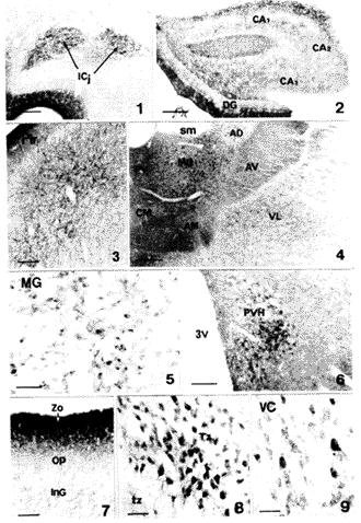
图1 示Calleja细胞岛内的mGluR7样免疫反应纤维。标尺示200μm
图2 示海马(CA1~CA3)和齿状回(DG)内的mGluR7样免疫反应细胞和纤维。标尺示350μm
, http://www.100md.com
图3 示梨状皮质内的mGluR7样免疫反应胞体和纤维。标尺示150μm
图4 示丘脑内的mGluR7样免疫反应细胞.AM.前内侧核 AD.前背侧核 AV.前腹侧核 CM.中央内侧核 MD.背内侧核 PV.丘脑室旁核 sm.丘脑髓纹 VL.腹外侧核 标尺示400μm
图5 示内侧膝状体(MG)内的mGluR7样免疫反应细胞和纤维.标尺示150μm
图6 示下丘脑室旁核(PVH)内的mGluR7样免疫反应细胞和纤维。3V.第三脑室 标尺示200μm
图7 示上丘内的mGluR7样免疫反应纤维。InG.中灰质层 OP.视纤维层 SuG.浅灰层 Zo.带状层 标尺示150μm
图8,9 示斜方体(Tz,图8)和腹侧耳蜗核(VC,图9)内的mGluR7样免疫反应细胞。标尺示75μm
, 百拇医药
Fig.1 Distribution of mGluR7-LI fibers in the islands of Calleja(ICj).Bar=200μmFig.2 Distribution of mGluR7-LI neuronal cell bodies and fibers in the hippocampus and dentate gyrus (DG).CA1-CA3,CA1-CA3 fields of the hippocampus.Bar=350μmFig.3 Distribution of mGluR7-LI neuronal cell bodies and fibers in the piriform cortex.Bar=150μmFig.4 Distribution of mGluR7-LI neurons in the thalamus.AD:anterodorsal thalamic nucleus;AM:anteromedial thalamic nucleus;AV:anteroventral thalamic nucleus;CM:central medial thalamic nucleus; MD:mediodorsal thalamic nucleus; sm:stria medullaris thalamus; VL:ventrolateral thalamic nucleus. Bar=400μmFig.5 Distribution of mGluR7-LI neuronal cell bodies and fibers in the medial geniculate body (MG).Bar=150μmFig.6 Distribution of mGluR7-LI neuronal cell bodies and fibers in the paraventricular hypothalamic nucleus(PVH).3V,third ventricle.Bar=200μmFig.7 Distribution of mGluR7-LI fibers in the superior colliculus.InG:intermediate white layer;OP:optic nerve layer; SuG:superficial gray layer; Zo,zonal layer.Bar=150μmFigs.8,9 Distribution of mGluR7-LI neuronal cell bodies in the trapezoid body(Tz,Fig.8)and ventral cochlear nucleus(VC,Fig.9).Bar=75μm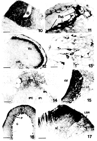
, http://www.100md.com
图10,11 示孤束核(Sol,图10)和三叉旁核(Pa5,图11)内的mGluR7样免疫反应纤维。*.血管 标尺示150μm
图12 示三叉神经尾侧亚核(Vc)延髓网状结构(LRt)内的mGluR7样免疫反应细胞和纤维。t.三叉脊束 标尺示350μm
图13 示中缝大核内的mGluR7样免疫反应细胞。标尺示75μm
图14 示脚间核内的mGluR7样免疫反应细胞和纤维。IPC.尾侧亚核 IPI.中间亚核 IPL.外侧亚核 IPR.吻侧亚核 标尺示150μm
图15 示蓝斑(LC)内的mGluR7样免疫反应细胞和纤维。4V.第四脑室 标尺示150μm
图16 示小脑皮质内的mGluR7样免疫反应细胞和纤维。G.颗粒层 M.分子层 P.蒲肯野细胞层 标尺示150μm
, http://www.100md.com
图17 示脊髓背角内的mGluR7样免疫反应细胞和纤维。Ⅰ~Ⅳ.Ⅰ~Ⅳ层 LSN.外侧脊核 标尺示150μm
Figs.10,11 Distribution of mGluR7-LI fibers in the nucleus of the solitary tract (Sol,Fig.10)and paratrigeminal nucleus(Pa5).*:blood vessel.Bar=150μm Fig.12 Distribution of mGluR7-LI neuronal cell bodies and fibers in the caudal spinal trigeminal nucleus(Vc)and lateral medullary reticular formation(LRt).t:spinal trigeminal tract.Bar=350μmFig.13 Distribution of mGluR7-LI neuronal cell bodies in the magnus raphe nucleus.Bar=75μmFig.14 Distribution of mGluR7-LI neuronal cell bodies and fibers in the interpeduncular.IPC:caudal subnucleus;IPI:intermediate subnucleus; IPL:lateral subnucleus; IPR:rostral subnucleus.Bar=150μmFig.15 Distribution of mGluR7-LI neuronal cell bodies and fibers in the locus coeruleus(LC).4V:fourth ventricle.Bar=150μmFig.16 Distribution of mGluR7-LI neuronal cell bodies and fibers in the cerebellar cortex.G:granular layer;M:molecular layer;P:Purkinje cell layer.Bar=150μmFig.17 Distribution of mGluR7-LI neuronal cell bodies and fibers in the dorsal horn of the spinal cord.Ⅰ,Ⅱ,Ⅲ and Ⅳ:laminae Ⅰ-Ⅳ;LSN:lateral spinal nucleus.Bar=150μm
, 百拇医药
△ Department of Anatomy and K.K.Leung Brain Research Center,The Fourth Military Medical University,Xi′an 710032,China
参考文献
[1]Nakanishi S.Molecular diversity of glutamate receptors and implications for brain function Science,1992,258(3):597
[2]Masu M,Tanabe Y,Tsuchida R,et al.Sequence and expression of a metabotropic glutamate receptor.Nature,1991,349(4):760
, 百拇医药 [3]Houamed KM,Kuijper TL,Gilbert BA,et al.Cloning,expression,and gene structure of a G protein-coupled glutamate receptor from rat brain,Science,1991,252(7):1318
[4]Tanabe Y,Masu M,Ishii,T et al.A family of metabotropic glutamate receptors.Neurons,1992,252(1):169
[5]Saugstad JA,Kinzie JM,Mulvihill ER,et al.Cloning and expression of a new member of the L-2-amino-4-phosphaonobutyric acid-sensitive class of metabotropic glutamate receptors.Mol Pharmacol,1994,45(2):367
, http://www.100md.com
[6]Nakajima Y,Iwakabe H,Akazawa C,et al.Molecular characterization of a novel retinal metabotropic glutamate receptor mGluR6 with a high agonist selectivity for L-2-amino-4-phosphonobutyrate.J Biol Chem,1993,268(16):11868
[7]Okamoto N,Hori S,Akazawa C,et al.Molecular characterization of a new metabotropic glutamate receptor mGluR7 coupled to inhibitory cyclic AMP signal transduction.J Biol Chem,1994,267(5):1231
[8]Pin JP,Dnvoisin R.The metabotropic glutamate receptors:structure and function.Neuropharmacololgy,1995,34(1):1
, 百拇医药
[9]Ohishi H.Akazawa C,Shigemoto R,et al.Distributions of the mRNA for L-2-amino-4-phosphonobutyrate-sentive metabotropic glutamate receptors,mGluR4 and mGluR7,in the rat brain,J Comp Neurol,1995,360(2):555
[10]Hearn TJ,Ganong AH,Cotman CW.Antagonism of lateral olfactory tract synaptic potentials in rat prepyriform cortex slices.Brain Res,1986,379(2):372
[11]Hasselmo ME,Bower JM.Selective suppression of afferent but not intrinsic fiber synaptic transmission by 2-amino-4-phosphonobutyric acid(AP4)in piriform cortex.Brain Res,1991,548(2):248
, 百拇医药
[12]Baskys A,Malenka RC.Agonists at metabotropic glutamate receptors presynaptically inhibit EPSPs in neonatal rat hippocampus.J Physiol,1991,444(2):687
[13]Ohishi H,Nomura S,Ding YQ,et al.Presynaptic Localization of a metabotropic glutamate receptor,mGluR7,in the primary afferent neurons:an immuno histo chemical study in the rat.Neurosci Lett,1995,202(1):85
[14]Shigemoto R,Kinoshita A,Wada E,et al.Differential presynaptic localization of metabotropic glutamate recerptor subtypes in the rat hippocampus.J Neurosci,1997,17(19):7503
[15]Li JL,Ohishi H,Kaneko T et al.Immunohistochemical localization of a metabotropic glutamate receptor,mGluR7,in ganglion neurons of the rat:with special reference to the presence in glutamatergic ganglion neurons.Neurosci Lett.1996,204(1):9
收稿1997-12 修回1998-04, http://www.100md.com
单位:李金莲 丁玉强 李继硕第四军医大学解剖学教研室,梁銶琚脑研究中心,西安 710032大石 仁重本隆一水野 升 日本京都大学医学部高级脑形态学研究室,京都 606
关键词:代谢型谷氨酸受体7亚型;谷氨酸;脑;脊髓;免疫细胞化学;大鼠;
解剖学报990101
【摘要】 目的 为研究代谢型谷氨酸受体在中枢神经系统内的功能作用提供形态学依据。 方法 用免疫组织化学技术,在光学显微镜下观察代谢型谷氨酸受体7亚型(mGluR7)在大鼠脑和脊髓内的分布。 结果 mGluR7阳性浓染的神经元胞体和纤维主要密集分布于Calleja岛、海马、齿状回、内侧缰核、橄榄顶盖前核、上丘带状层、三叉神经尾侧亚核浅层、三叉旁核、中缝大核、中缝苍白核、蓝斑、脊髓背角浅层和外侧脊核。呈中等强度染色的神经元胞体和纤维见于嗅结节、前嗅核、梨状皮质、隔伞核、终纹床核、下丘脑外侧区、下丘脑室旁核、乳头体上核、乳头体内、外核、丘脑大部分核团、内外侧膝状体、视束核、红核、黑质、脚间核、桥核、臂旁外侧核、上橄榄复合体、斜方体核、三叉神经运动核、面神经核、疑核、耳蜗核、前庭核簇、楔束外核、孤束核、舌下神经核、舌下神经前置核、中缝隐核、延髓网状结构、小脑蒲肯野细胞层、脊髓中间带外侧核、Onuf核和中央管周围灰质。此外,在许多脑区还可见淡染的和散在分布的阳性胞体和纤维。 结论 mGluR7广泛分布于大鼠中枢神经系统内,提示它在谷氨酸引起的兴奋性信息传递中可能起着重要作用。
, http://www.100md.com
IMMUNOHISTOCHEMICAL LOCALIZATION OF A METABOTROPIC GLUTAMATERECEPTOR,mGLUR7,IN THE CENTRAL
NERVOUS SYSTEM OF THE RAT
Li Jinlian△, Ding Yuqiang, Li Jishuo, Ohishi Hitoshi*, Shigemoto Ryuichi*,Mizuno Noboru*
(Department of Anatomy and K.K.Leung Brain Research Center,The Fourth Military Medical
University,Xi′an;*Department of Morphological Brain Science,Faculty of Medicine,Kyoto University,Kyoto,Japan)
, http://www.100md.com
【Abstract】 Objective In order to study possible functions of the metabotropic glutamate receptors in the central nervous system,the localization of metabotropic glutamate receptor 7 subtype in the brain and spinal cord of the rat was examined.Methods Immunohistochemical staining techniques were used,and immunostained sections were observed under a light microscopy.Results Many neuronal cell bodies and fibers with dense mGluR7-like immunoreactivity(-LI) were intensely distributed in the island of Calleja,hippocampus,dentate gyrus,medial habenular nucleus,olivary pretectal nucleus,zonal layer of the superior colliculus,superficial layers of the caudal spinal trigeminal nucleus,paratrigeminal nucleus,raphe magnus nucleus,raphe nucleus pallidus,locus coeruleus,superficial layers and lateral spinal nucleus of the spinal cord.Neuronal cell bodies and fibers with moderate mGluR7-LI were seen in the olfactory bulb,anterior olfactory nucleus,piriform cortex,septofimbrial nucleus,bed nucleus of the strial terminalis,lateral hypothalamic area,paraventricular hypothalamic nucleus,supramammillary nucleus,medial and lateral mammillary nuclei,most part of the thalamus,medial and lateral geniculate bodies,nucleus of the optic tract,red nucleus,substantia nigra,interpeduncular nucleus,pontine nuclei,lateral parabrachial nucleus,superior olivary complex,nucleus of the trapezoid body,motor nucleus of the trigeminal nerve,facial nucleus,ambiguus nucleus,cochlear nucleus,vestibular nuclei,nucleus of the solitary tract,hypoglossal nucleus,prepositus hypoglossal nucleus,medullary reticular formation,Purkinje cells of the cerebellum,nucleus raphe obscurus,intermediolateral nucleus of the spinal cord,Onuf′s nucleus and lamina X of the spinal cord.In addition,weak mGluR7-LI was detected in some other regions of the brain and spinal cord.Conclusion The present results indicate that mGluR7 is widely distributed in the central nervous system of the rat,and suggest that mGluR7 might play an important role in the excitatory synaptic transmission induced by glutamate.
, http://www.100md.com
【Key words】 Metabotropic glutamate receptor 7 subtype; Glutamate; Brain; Spinal cord; Immunohistochemistry; Rat
众所周知,谷氨酸(Glu)是广泛存在于哺乳动物中枢神经系统内的一种兴奋性神经递质,与快速兴奋性突触传递、神经元的发育和死亡、突触的可塑性以及某些神经疾病的发病密切相关[1]。长期以来人们一直认为Glu是通过激活了配体门控性阳离子通道而发挥作用,此类受体被称为离子型谷氨酸受体。近年来,大量生理学和药理学实验表明,Glu还可通过与G蛋白偶联的一类受体来调节兴奋性突触的活动和神经元的兴奋性,此类受体被命名为代谢型谷氨酸受体(metabotropic glutamate receptors,mGluRs)。目前应用分子生物学技术已成功地克隆出mGluR的8种亚型(mGluR1-8)[2~7]。按照各亚型间氨基酸序列的同源性和偶联的细胞内第二信使及对激动剂的选择性,将mGluR1-8分为3组:(1)mGluR1和mGluR5;(2)mGluR2和mGluR3;(3)mGluR4、mGluR6、mGluR7和mGluR8。第3组的特征是可以抑制细胞内腺苷酸环化酶的活性使细胞内cAMP的含量降低,并对激动剂L-2氨基-4-磷酰丁酸(1-2-amino-4-phosphonobutyrate,L-AP4)具有高度亲合力[8]。这组受体中的mGluR7为含874个氨基酸残基膜蛋白,并具有G蛋白偶联受体的7次跨膜结构特征[7]。原位杂交组织化学研究发现mGluR7广泛分布在大鼠中枢神经系统内[9]。但原位杂交组织化学只能用于研究神经元是否表达mGluR7 mRNA,不能用于受体蛋白的定位研究。而受体蛋白的定位研究对了解受体的功能是不可缺少的。此外,已有的药理学研究表明,mGluR7在一些脑区可作为自受体通过突触前机制抑制谷氨酸的释放[10~12]。因此,本研究采用免疫组织化学方法系统地观察了mGluR7在大鼠脑和脊髓内分布状况。
, 百拇医药
材料和方法
雄性Wistar大鼠5只,体重250~300g。在腹腔内注射戊巴比妥钠的深麻状态下,开胸经左心室插管,先以150ml 0.02 mol/L的磷酸盐缓冲液(PBS,pH7.3)冲洗血液,再用500ml含4%多聚甲醛和1%苦味酸的0.1mol/L的磷酸缓冲液(PB,pH 7.3)灌注固定1h。立即取出脑和脊髓,置于含30%蔗糖的PB内过夜(4℃)。冰冻连续切片(厚30μm)。切片分为3套,收集于PBS内。第1套切片用于mGluR7免疫组织化学染色,具体步骤如下:切片置于兔抗mGluR7血清(0.25mg/L)[13]内室温下孵育过夜,然后移入结合生物素的羊抗兔血清(1∶200,Vector)室温下孵育2h,最后在ABC复合物内反应2h。DAB和H2O2呈色后裱片、脱水透明、DPX封片。第2组切片用于对照实验。一是用过量的抗原(GST-mGluR7融合蛋白,内含mGluR7 C-末端47个氨基酸残基)与mGluR7抗体室温下预孵2h,按上述步骤进行染色,二是用正常兔血清替代一抗,再按上述步骤进行染色。第3套切片经Nissl染色后用于确定核团的名称和位置。
, 百拇医药
结果
在进行吸收实验和替代实验的脑和脊髓的切片上,均未见免疫反应产物。
免疫组织化学染色结果表明,mGluR7样免疫反应成分广泛分布于大鼠脑和脊髓内,为便于描述,将mGluR7阳性产物的强度相对地分为浓(+++,如蓝斑,图15)、中(++,如下丘脑室旁核,图6)和淡(+,如丘脑腹外侧核,图4)3个等级。按此标准,将mGluR7样免疫反应产物的分布归纳成附表。星号表示该部位的神经元的胞体亦为阳性。
1. 嗅系统和大脑皮质区
主嗅球和副嗅球的僧帽细胞、嗅结节、Calleja岛(图1)、海马分子层和多形层、齿状回和梨状皮质内可见中等密度以上的mGluR7阳性神经元和纤维(图2)。前嗅核、大脑皮质、梨状皮质(图3)、扣带回皮质、压部后皮质、嗅周皮质、内嗅皮质、旁下托、前下托内可见淡染且呈散在分布的阳性神经元胞体和纤维。
, 百拇医药
2. 前脑的皮质下区
隔伞核、终纹床核及丘脑的部分核团(图4),如内侧缰核、前背侧核、丘脑内侧背核、丘脑中央外侧核、丘脑腹后内侧核和外侧核、丘脑胶状质核、丘脑后核群、内侧膝状体(图5)和外侧膝状体内可见大量中等强度染色的mGluR7阳性神经元的胞体和少量阳性纤维。此外,下丘脑室旁核(图6)、下丘脑外侧区、乳头体上核、乳头体内侧核和外侧核也含有一定数量中等强度染色的mGluR7阳性神经元的胞体和阳性纤维。
附表 mGluR7免疫反应产物在大鼠中枢神经系统内的分布
Table Distribution of mGluR7-LI in the central nervous system of the rat 观察区域
observed regions
, http://www.100md.com 强度
intensity
观察区域
observed regions
强度
intensity
嗅觉系统(olfactory system)
内侧杏仁核(medial amygdaloid nucleus)
+*
主嗅球(main olfactory bulb)
其他(other nuclei)
, 百拇医药
-
僧帽细胞(mitral cell)
++*
视前区和下丘脑(preoptic region and hypothalamus)
其他(other layers)
-
外侧视前区(lateral preoptic area)
+
副嗅球(accessory olfactory bulb)
内侧视前区(medial preoptic area)
, http://www.100md.com
+
僧帽细胞(mitral cell)
++*
内侧视前核(medial preoptic nucleus)
+*
其他(other layers)
-
视交叉上核(suprachiasmatic nucleus)
-
前嗅核(anterior nucleus)
, http://www.100md.com ++*
视上核(supraoptic nucleus)
+*
嗅结节(olfactory tubercle)
+
下丘脑前区(anterior hypothalamus)
+
Calleja(islands of Calleja)
+++
下丘脑外侧区(lateral hypothalamic area)
, 百拇医药
++*
大脑皮质(cerebral cortex)
室周核(periventricular nucleus)
+*
Ⅰ层(layer Ⅰ)
+
室旁核(paraventricular nucleus)
++*
Ⅱ~Ⅳ层(layers Ⅱ-Ⅳ)
+*
, http://www.100md.com
弓状核(arcuate nucleus)
+*
Ⅴ~Ⅵ层(layers Ⅴ-Ⅵ)
+
下丘脑背侧区(dorsal hypothalamic area)
+*
边缘皮质(limbic cortex)
下丘脑腹内侧核(ventromedial hypothalamic nucleus)
+*
梨状皮质(piriform cortex)
, http://www.100md.com
++*
下丘脑后区(posterior hypothalamic area)
+*
扣带皮质(cingulate cortex)
+*
乳头体前核(premammillary nucleus)
-
压后皮质(retrosplenial cortex)
+*
乳头体上核(supramammillary nucleus)
, 百拇医药
++*
嗅周皮质(perihinal cortex)
+*
乳头体内侧核(medial mammillary nucleus)
++*
内嗅皮质(entorhinal cortex)
+*
乳头体外侧核(lateral mammillary nucleus)
++*
, 百拇医药 旁下托(parasubcular cortex)
+*
丘脑上部和底部(epithalamus and subthalamus)
前下托(presubicular cortex)
+*
内侧缰核(medial habenular nucleus)
+++*
海马结构(hippocampus)
外侧缰核(lateral habenular nucleus)
, http://www.100md.com
-
齿状回(dentate gyrus)
+++*
底丘脑核(subthalamic nucleus)
+*
分子层(molecular layer)
+++*
未定带(zona incerta)
+
颗粒细胞层(graule cell layer)
-
, 百拇医药
丘脑(thalamus)
多形层(polymorph layer)
++
丘脑网状核(thalamic reticular nucleus)
+
CA1~CA3区(CA1-CA3)
其他核团(other nuclei)
+~++
分子层(molecular layer)
+++*
, 百拇医药 脑干(bain stem)
颗粒细胞层(graule cell layer)
-
顶盖前区(pretectal region)
多形层(polymorph layer)
++*
橄榄顶盖前核(olivary pretectal nucleus)
+++
隔区(septal region)
视束核(nucleus of the optic tract)
, http://www.100md.com
++
内侧隔核(medial septal nucleus)
+
其他区域(other regions)
-
外侧隔核(lateral septal nucleus)
-
上丘(superior colliculus)
隔伞核(septofimbrial nucleus)
++*
, 百拇医药 带状层(zonal layer)
+++
斜角带(diagonal band)
+
浅灰层(superficial gray layer)
++
终纹床核(bed nucleus of the stria strminalis)
++*
视神经层~中白层(optic nerve-intermediate white layers)
+
, http://www.100md.com
基底神经节(basal ganglia)
深灰层(deep gray layer)
+*
尾壳核(daudate putamen)
+
深白层(deep white layer)
+
苍白球(globus pallidum)
+*
下丘(inferior colliculus)
, 百拇医药
+~++*
屏状核(claustrum)
+
外侧丘系核(lateral lemniscus nuclei)
+*
伏核(accumbens nucleus)
+
上橄榄复合体(superior olivary complex)
++*
腹侧苍白球(ventral pallidum)
, http://www.100md.com
+*
斜方体核(nucleus of the trapezoid body)
++*
脚内核(entopeduncular nucleus)
+*
耳蜗核(cochlear nuclei)
++*
杏仁核簇(amygdloid complex)
前庭核簇(vestibular nuclei)
, 百拇医药
外侧杏仁核(lateral amygdaloid nucleus)
+
前庭内侧核(medial vestibular nucleus)
++*
前庭外侧核(lateral vestibular nucleus)
+*
吻侧亚核(rostral subnucleus)
+++
前庭上核(superior vestibular nucleus)
, 百拇医药
+*
尾侧亚核(caudal subnucleus)
+
前庭脊核(spinal vestibular nucleus)
++*
背外侧亚核(dorsolateral subnucleus)
++
三叉神经感觉核簇(trigeminal sensory complex)
中间亚核(intermediate subnucleus)
, 百拇医药 +*
三叉神经中脑核(mesencephalic trigeminal nucleus)
-
外侧亚核(lateral subnucleus)
++
三叉神经感觉主核(principal sensory trigeminal nucleus)
+*
楔形核(cuneiform nucleus)
+
三叉神经脊束核(spinal trigeminal nucleus)
, 百拇医药
臂旁外侧核(lateral parabrachial nucleus)
++*
吻侧亚核(oral subnucleus)
+*
臂旁内侧核(medial parabrachial nucleus)
+*
极间亚核(interpolar subnucleus)
+*
蓝斑(locus coeruleus)
, 百拇医药
+++*
尾侧亚核(caudal subnucleus)
KF核(
-Fuse nucleus)+*
Ⅰ~Ⅱ层(laminae Ⅰ and Ⅱ)
+++
被盖背侧核(dorsal tegmental nucleus)
+
Ⅲ~Ⅳ层(laminae Ⅲ and Ⅳ)
, 百拇医药
+*
被盖背外侧核(laterodorsal tegmental nucleus)
+
孤束核(nucleus of the solitary tract)
++~+++
舌下神经前置核(prepositus nucleus)
++
三叉旁核(paratrigeminal nucleus)
+++
中脑网状结构(mesencephalic reticular formation)
, 百拇医药
+*
楔束外核(external cuneate nucleus)
++*
桥脑网状结构(pontine reticular formation)
+*
薄束核(gracile nucleus)
+*
延髓网状结构(medullary reticular formation)
++*
, http://www.100md.com
小脑前核(precerebellar nuclei)
+
巨细胞网状核(gigantocellular reticular nucleus)
+*
桥核(pontine nuclei)
++*
线形中缝核(linear raphe nucleus)
+
下橄榄核(inferior olivary complex)
+*
, 百拇医药
正中中缝核(median raphe nucleus)
+*
外侧网状核(lateral reticular nucleus)
+*
中缝背核(dorsal raphe nucleus)
+*
运动及运动有关核团(motor and motor-related nuclei)
中缝大核(magnus raphe nucleus)
+++*
, http://www.100md.com
红核(red nucleus)
++*
中缝苍白核(raphe nucleus pallidus)
+++*
黑质(substantia nigra)
++
中缝隐核(raphe nucleus obscurus)
++*
动眼神经核(oculomotor nucleus)
+*
, http://www.100md.com
最后区(area postrma)
+
滑车神经核(abducens nucleus)
+*
小脑(cerebellum)
E-W核(Edinger-Westphal nucleus)
+*
小脑皮质(cerebellar cortex)
Darkschewitsch核(Darkschewitsch nucleus)
+*
, http://www.100md.com
分子层(molecular layer)
++
Cajal中介核(interstitial nucleus of Cajal)
+*
蒲肯野细胞层(Purkinje cell layer)
++*
三叉神经运动核(motor nucleus of the trigeminal nerve)
++*
颗粒层(granular layer)
, http://www.100md.com
-
面神经核(facial nucleus)
++*
小脑深核(deep cerebellar nuclei)
++*
疑核(ambiguus nucleus)
++*
脊髓(spinal cord)
迷走神经背运动核(dorsal motor nucleus of
Ⅰ~Ⅱ层(laminae Ⅰ and Ⅱ)
, http://www.100md.com
+++
the vagus nerve)
+
Ⅲ~Ⅷ层(laminae Ⅲ-Ⅷ)
+
舌下神经核(hypoglossal nucleus)
++*
Ⅹ层(lamina Ⅹ)
++
其他(others)
前角运动神经元(motor neurons)
, 百拇医药
+*
中脑导水管周围灰质(periaqueductal gray)
-
Onuf核(Onuf′s nucleus)
++*
脚间核(interpeduncular nucleus)
mGluR7样免疫反应产物相对强度:+++:浓,++:中,+:淡,-:阴性。*表示该核团内含阳性胞体
Relative intensity of mGluR7-LI.+++:high; ++:moderate; +:low; -:negative.*Areas where neuronal cell bodies were detected.
, 百拇医药
含有稀疏且淡染的胞体和纤维的区域有:内外侧隔核、伏核、隔三角核、斜角带核、腹侧苍白球、苍白球、屏状核、尾壳核、杏仁内侧核和外侧核、内侧和外侧视前区、视上核、下丘脑前区、室周核、弓状核、下丘脑背侧区、下丘脑腹内侧核、下丘脑后区和丘脑的大部分核团,包括前腹侧核、前内侧核、中央正中核、外侧背核、腹外侧核、连结核、菱形核、束旁核、网状核、未定带和底丘脑核等。
3. 脑干
顶盖前区的橄榄顶盖前核和视束核内可见密集的mGluR7阳性纤维。在与视听觉有关的脑区如上丘(图7)、外侧丘系核、上橄榄复合体、斜方体核(图8)、耳蜗核(图9)内观察到mGluR7的阳性胞体和纤维。在其他感觉核团中也发现有mGluR7阳性胞体和纤维,如前庭核簇、孤束核(图10)、楔束外核、三叉旁核(图11)、三叉神经感觉主核、三叉神经脊束核吻侧、极间亚核和尾侧亚核的浅层(图12)以及后索核。在运动或运动相关核团如红核、黑质、面神经核、三叉神经运动核、E-W核、动眼神经核、滑车神经核、外展神经核、舌下神经核、迷走神经背核和疑核内也观察到mGluR7阳性的胞体和纤维。
, 百拇医药
脑干的其他部位如中缝大核(图13)、脚间核(图14)、蓝斑(图15)、桥核、中缝苍白核、中缝隐核、舌下神经前置核和延髓网状结构内分布有中等密度以上的mGluR7阳性胞体和纤维。而在脑桥网状被盖核、下橄榄核簇、臂旁内侧核、KF核、脑干被盖核和最后区等处仅见到浅染或散在分布的胞体和纤维。
4. 小脑和脊髓
在小脑皮质的蒲肯野细胞的胞体呈阳性反应、分子层内分布有密集的mGluR7阳性纤维(图16),小脑深部核团的神经元呈中等强度的免疫反应。
在脊髓,浓密的mGluR7阳性纤维和终末主要分布于背角Ⅰ、Ⅱ层,其中Ⅰ层和Ⅱ层的外侧部更为浓密(图17)。但在Ⅰ层和Ⅱ层未见阳性的胞体。此外,外侧脊核内分布有深染的阳性胞体和致密的纤维。而在胸髓的后角深层、中间带外侧核和腰骶髓的Onuf核内只见到少量mGluR7阳性胞体和纤维。
, http://www.100md.com 讨论
本研究采用免疫组织化学方法观察发现mGluR7样免疫反应产物广泛分布于大鼠中枢神经系统内,这一结果与Ohishi等[9]用原位杂交组织化学方法所报道的mGluR7 mRNA的分布基本一致,尤其在嗅球、腹侧苍白球、海马、纹状体、大脑皮质、丘脑、红核、黑质、上丘、脚间核、桥核、外侧丘系核、耳蜗核、前庭核、楔束外核、耳蜗核、延髓网状结构等脑区。本研究所使用的抗mGluR7抗体经Western blotting实验证实了其特异性,即只与表达mGluR7的中国苍鼠卵细胞(CHO)的膜提取物结合,而与表达其他代谢型谷氨酸受体的CHO膜提取物之间无任何反应,且该抗体识别的脑组织中的蛋白的分子量与克隆的mGluR7的分子量一致[13]。此外,抗体吸收实验和替代实验也进一步证实该抗体的特异性。因此,本实验的结果是可靠的。
但是,在一些脑区原位杂交的结果与本研究的结果间存在差异。如在Calleja岛、内侧缰核、乳头体外侧核、红核、面神经核、小脑深核仅观察到少量阳性神经元表达mGluR7 mRNA[9],而本研究免疫组织化学的结果显示,这些区域内有相当数量的神经元呈现免疫反应性并含有一定数量的阳性纤维。相反,本研究在外侧隔核、隔三角核、斜角带核、杏仁基底内外侧核、杏仁中央核、视交叉上核、内侧视前区、下丘脑前区、外侧缰核、导水管周围灰质等部位仅见到很少或未观察到阳性神经元,而Ohishi等[9]在上述部位观察到大量的mGluR7 mRNA阳性神经元。这些差异可能是由于方法学的不同引起的。首先,原位杂交显示的是胞体内的mRNA,而免疫组织化学则是显示胞体和突起内的mGluR7蛋白。已有大量药理学研究表明,在梨状皮质、海马等处,mGluR7可通过突触前机制抑制轴突终末释放谷氨酸[10~12],提示mGluR7在胞体合成后可能被运输到轴突内而发挥作用。这可能是两种研究方法的结果存在差异的原因之一。此外,任何一种免疫组织化学染色产物都不可能只定位于所要探查的成分内,即不能排除制备的抗体与组织内和目标抗原结构相似的蛋白结合。最后,需要考虑到的是所制备的抗体未能与组织内所有的mGluR7结合,因为所制备的抗体只识别mGluR7整个分子的一部分,在不同脑区其空间结构可能存在差异从而使其抗原性发生变化。
, 百拇医药
如前所述,L-AP4是第3组mGluRs的特异性激动剂。以往的研究发现在一些脑区L-AP4对谷氨酸能终末释放谷氨酸具有抑制作用,即第3组mGluRs常作为分布于突触前的自受体而发挥作用。如嗅球僧帽细胞发出的经外侧嗅束投射到梨状皮质的轴突终末内含有谷氨酸,而L-AP4可通过突触前机制抑制谷氨酸的释放[10,11]。此外,在海马和齿状回,由内嗅皮质发出的向齿状回的谷氨酸能投射纤维和海马CA3区的锥体细胞发出的向CA1投射的含谷氨酸的轴突侧支均为L-AP4的作用部位[12]。本研究发现在上述脑区分布有mGluR7样免疫反应纤维,这些阳性纤维很可能是含mGluR7的阳性轴突及其终末。事实上,最近的免疫电镜研究已证实海马内分布有大量的mGluR7阳性的轴突终末[14]。
谷氨酸作为一种重要的兴奋性神经递质在伤害性信息的传递中起着重要作用。我们以往的研究发现,背根节和三叉神经节内分布有大量的mGluR7阳性胞体,以中小型居多,且均为磷酸激活的谷氨酰胺酶(谷氨酸的合成酶)阳性[15]。切断背根后,背角浅层(Ⅰ~Ⅱ层)内mGluR7样免疫反应产物近乎完全消失,免疫电镜研究证实背角浅层内mGluR7阳性成分多为阳性的轴突终末[13]。因此,有理由认为mGluR7在谷氨酸介导的伤害性信息传递中起着重要的作用,即可能作为自受体来调节伤害性刺激引起的初级传入终末谷氨酸的释放量,这种调节可能与伤害性刺激引起的动物行为反应相一致。此问题有待于进一步研究。
, http://www.100md.com
需要指出的是在大鼠脑和脊髓的很多区域分布有mGluR7阳性胞体。这些脑区包括非谷氨酸能的细胞,如小脑含GABA的蒲肯野细胞和脑干内一些胆碱能的运动核团。在这些部位mGluR7可能作为突触后受体而发挥作用。
综上所述,广泛分布于大鼠中枢神经系统内的mGluR7在介导谷氨酸引起的兴奋性突触传递中起着重要作用,尤其是其作为自受体调节轴突终末谷氨酸的释放是今后一个值得深入探讨的问题。
本文的图片工作得到了原悦萍同志的帮助,谨致谢意。
图版说明
图1 示Calleja细胞岛内的mGluR7样免疫反应纤维。标尺示200μm
图2 示海马(CA1~CA3)和齿状回(DG)内的mGluR7样免疫反应细胞和纤维。标尺示350μm
, http://www.100md.com
图3 示梨状皮质内的mGluR7样免疫反应胞体和纤维。标尺示150μm
图4 示丘脑内的mGluR7样免疫反应细胞.AM.前内侧核 AD.前背侧核 AV.前腹侧核 CM.中央内侧核 MD.背内侧核 PV.丘脑室旁核 sm.丘脑髓纹 VL.腹外侧核 标尺示400μm
图5 示内侧膝状体(MG)内的mGluR7样免疫反应细胞和纤维.标尺示150μm
图6 示下丘脑室旁核(PVH)内的mGluR7样免疫反应细胞和纤维。3V.第三脑室 标尺示200μm
图7 示上丘内的mGluR7样免疫反应纤维。InG.中灰质层 OP.视纤维层 SuG.浅灰层 Zo.带状层 标尺示150μm
图8,9 示斜方体(Tz,图8)和腹侧耳蜗核(VC,图9)内的mGluR7样免疫反应细胞。标尺示75μm
, 百拇医药
Fig.1 Distribution of mGluR7-LI fibers in the islands of Calleja(ICj).Bar=200μmFig.2 Distribution of mGluR7-LI neuronal cell bodies and fibers in the hippocampus and dentate gyrus (DG).CA1-CA3,CA1-CA3 fields of the hippocampus.Bar=350μmFig.3 Distribution of mGluR7-LI neuronal cell bodies and fibers in the piriform cortex.Bar=150μmFig.4 Distribution of mGluR7-LI neurons in the thalamus.AD:anterodorsal thalamic nucleus;AM:anteromedial thalamic nucleus;AV:anteroventral thalamic nucleus;CM:central medial thalamic nucleus; MD:mediodorsal thalamic nucleus; sm:stria medullaris thalamus; VL:ventrolateral thalamic nucleus. Bar=400μmFig.5 Distribution of mGluR7-LI neuronal cell bodies and fibers in the medial geniculate body (MG).Bar=150μmFig.6 Distribution of mGluR7-LI neuronal cell bodies and fibers in the paraventricular hypothalamic nucleus(PVH).3V,third ventricle.Bar=200μmFig.7 Distribution of mGluR7-LI fibers in the superior colliculus.InG:intermediate white layer;OP:optic nerve layer; SuG:superficial gray layer; Zo,zonal layer.Bar=150μmFigs.8,9 Distribution of mGluR7-LI neuronal cell bodies in the trapezoid body(Tz,Fig.8)and ventral cochlear nucleus(VC,Fig.9).Bar=75μm
, http://www.100md.com
图10,11 示孤束核(Sol,图10)和三叉旁核(Pa5,图11)内的mGluR7样免疫反应纤维。*.血管 标尺示150μm
图12 示三叉神经尾侧亚核(Vc)延髓网状结构(LRt)内的mGluR7样免疫反应细胞和纤维。t.三叉脊束 标尺示350μm
图13 示中缝大核内的mGluR7样免疫反应细胞。标尺示75μm
图14 示脚间核内的mGluR7样免疫反应细胞和纤维。IPC.尾侧亚核 IPI.中间亚核 IPL.外侧亚核 IPR.吻侧亚核 标尺示150μm
图15 示蓝斑(LC)内的mGluR7样免疫反应细胞和纤维。4V.第四脑室 标尺示150μm
图16 示小脑皮质内的mGluR7样免疫反应细胞和纤维。G.颗粒层 M.分子层 P.蒲肯野细胞层 标尺示150μm
, http://www.100md.com
图17 示脊髓背角内的mGluR7样免疫反应细胞和纤维。Ⅰ~Ⅳ.Ⅰ~Ⅳ层 LSN.外侧脊核 标尺示150μm
Figs.10,11 Distribution of mGluR7-LI fibers in the nucleus of the solitary tract (Sol,Fig.10)and paratrigeminal nucleus(Pa5).*:blood vessel.Bar=150μm Fig.12 Distribution of mGluR7-LI neuronal cell bodies and fibers in the caudal spinal trigeminal nucleus(Vc)and lateral medullary reticular formation(LRt).t:spinal trigeminal tract.Bar=350μmFig.13 Distribution of mGluR7-LI neuronal cell bodies in the magnus raphe nucleus.Bar=75μmFig.14 Distribution of mGluR7-LI neuronal cell bodies and fibers in the interpeduncular.IPC:caudal subnucleus;IPI:intermediate subnucleus; IPL:lateral subnucleus; IPR:rostral subnucleus.Bar=150μmFig.15 Distribution of mGluR7-LI neuronal cell bodies and fibers in the locus coeruleus(LC).4V:fourth ventricle.Bar=150μmFig.16 Distribution of mGluR7-LI neuronal cell bodies and fibers in the cerebellar cortex.G:granular layer;M:molecular layer;P:Purkinje cell layer.Bar=150μmFig.17 Distribution of mGluR7-LI neuronal cell bodies and fibers in the dorsal horn of the spinal cord.Ⅰ,Ⅱ,Ⅲ and Ⅳ:laminae Ⅰ-Ⅳ;LSN:lateral spinal nucleus.Bar=150μm
, 百拇医药
△ Department of Anatomy and K.K.Leung Brain Research Center,The Fourth Military Medical University,Xi′an 710032,China
参考文献
[1]Nakanishi S.Molecular diversity of glutamate receptors and implications for brain function Science,1992,258(3):597
[2]Masu M,Tanabe Y,Tsuchida R,et al.Sequence and expression of a metabotropic glutamate receptor.Nature,1991,349(4):760
, 百拇医药 [3]Houamed KM,Kuijper TL,Gilbert BA,et al.Cloning,expression,and gene structure of a G protein-coupled glutamate receptor from rat brain,Science,1991,252(7):1318
[4]Tanabe Y,Masu M,Ishii,T et al.A family of metabotropic glutamate receptors.Neurons,1992,252(1):169
[5]Saugstad JA,Kinzie JM,Mulvihill ER,et al.Cloning and expression of a new member of the L-2-amino-4-phosphaonobutyric acid-sensitive class of metabotropic glutamate receptors.Mol Pharmacol,1994,45(2):367
, http://www.100md.com
[6]Nakajima Y,Iwakabe H,Akazawa C,et al.Molecular characterization of a novel retinal metabotropic glutamate receptor mGluR6 with a high agonist selectivity for L-2-amino-4-phosphonobutyrate.J Biol Chem,1993,268(16):11868
[7]Okamoto N,Hori S,Akazawa C,et al.Molecular characterization of a new metabotropic glutamate receptor mGluR7 coupled to inhibitory cyclic AMP signal transduction.J Biol Chem,1994,267(5):1231
[8]Pin JP,Dnvoisin R.The metabotropic glutamate receptors:structure and function.Neuropharmacololgy,1995,34(1):1
, 百拇医药
[9]Ohishi H.Akazawa C,Shigemoto R,et al.Distributions of the mRNA for L-2-amino-4-phosphonobutyrate-sentive metabotropic glutamate receptors,mGluR4 and mGluR7,in the rat brain,J Comp Neurol,1995,360(2):555
[10]Hearn TJ,Ganong AH,Cotman CW.Antagonism of lateral olfactory tract synaptic potentials in rat prepyriform cortex slices.Brain Res,1986,379(2):372
[11]Hasselmo ME,Bower JM.Selective suppression of afferent but not intrinsic fiber synaptic transmission by 2-amino-4-phosphonobutyric acid(AP4)in piriform cortex.Brain Res,1991,548(2):248
, 百拇医药
[12]Baskys A,Malenka RC.Agonists at metabotropic glutamate receptors presynaptically inhibit EPSPs in neonatal rat hippocampus.J Physiol,1991,444(2):687
[13]Ohishi H,Nomura S,Ding YQ,et al.Presynaptic Localization of a metabotropic glutamate receptor,mGluR7,in the primary afferent neurons:an immuno histo chemical study in the rat.Neurosci Lett,1995,202(1):85
[14]Shigemoto R,Kinoshita A,Wada E,et al.Differential presynaptic localization of metabotropic glutamate recerptor subtypes in the rat hippocampus.J Neurosci,1997,17(19):7503
[15]Li JL,Ohishi H,Kaneko T et al.Immunohistochemical localization of a metabotropic glutamate receptor,mGluR7,in ganglion neurons of the rat:with special reference to the presence in glutamatergic ganglion neurons.Neurosci Lett.1996,204(1):9
收稿1997-12 修回1998-04, http://www.100md.com