卵巢成熟性囊性畸胎瘤恶变临床病理分析及人类乳头瘤病毒检测
作者:沈丹华 张雅贤 薛为成 颜婉嫦
单位:沈丹华 薛为成 北京医科大学人民医院病理科,北京 100044;颜婉嫦 张雅贤 香港大学玛丽医院
关键词:卵巢肿瘤;病理学;乳头状瘤病毒;人;分离和提纯;畸胎癌;病理
北京医科大学学报990414 摘 要 目的:分析卵巢成熟性囊性畸胎瘤恶变的临床病理学特征及其与人类乳头瘤病毒感染的关系。方法:回顾性研究了14例卵巢成熟性囊性畸胎瘤恶变临床及病理学特征,并且应用PCR技术对其中7例的瘤体组织及宫颈组织进行了人类乳头瘤病毒检测。结果:14例卵巢畸胎瘤恶变病例发病年龄为30~82岁(平均61.7岁),组织学类型包括鳞状细胞癌(11例)、腺癌、甲状腺乳头状腺癌及甲状腺类癌(各1例)。人类乳头瘤病毒检测没有发现阳性病例。结论:卵巢成熟性囊性畸胎瘤恶变是一种少见的病变,发病年龄以绝经后妇女为主,组织学类型以鳞状细胞癌最为多见,未发现HPV感染与其的关系。
, http://www.100md.com
学
中国图书资料分类法分类号 R737.31
Clinicopathological analysis and human papilloma virus status
of ovarian mature cystic teratoma with malignant transformation
SHEN Dan-Hua#, ZHANG Ya-Xian (CHEUNG A N Y), XUE Wei-Cheng, YAN Wan-Chang (NGAN H Y S)
(#Department of Pathology, People's Hospital, Beijing Medical University, Beijing 100044)
, 百拇医药
ABSTRACT Objective: To analyze the clinicopathological feature and status of human papilloma virus (HPV) infection of ovarian mature cystic teratoma with malignant transformation. Methods: The clinical and pathological data of 14 cases ovarian mature cystic teratoma with malignant transformation were analyzed. Detection of HPV in 7 cases of tumors and cervices was performed by PCR technique. Results: The ages of patients ranged from 30 to 82 years (mean, 61.7 years). The tumor size (diameter) ranged from 8 cm to 21 cm (mean, 13 cm). Histologically, there were 11 cases of squamous cell carcinoma (78.6%), one of adenocarcinoma, one of thyroid papillary carcinoma, and one of strumous carcinoid. There was no evidence of HPV infection in the cases studied. Conclusion: Ovarian mature cystic teratoma with malignant teratoma is uncommon and in most cases occurs in postmenopausal women. Squamous cell carcinoma is of the most common histological type. The absence of HPV suggests that there is no association between HPV infection and this tumour.
, http://www.100md.com
MeSH Ovarian neoplasms/pathol Papillomavirus, human/isol Teratocarcinoma/pathol
成熟性囊性畸胎瘤(mature cystic teratoma, MCT)是女性卵巢较常见的肿瘤,占所有卵巢肿瘤的10%~34%[1~3]。本病少数病例可以发生恶性转化,称为成熟性囊性畸胎瘤恶变(MCT with malignant transformation)。虽然畸胎瘤的3个胚层均可发生恶变,但鳞状细胞癌是最常见的组织类型。有许多研究显示人类乳头瘤病毒(human papilloma virus, HPV)感染与生殖系统的鳞状细胞癌有关。我们分析了14例来自北京和香港的卵巢MCT恶变患者的临床及病理学特征,并对其中7例鳞状细胞癌进行HPV检测,以探讨HPV感染与鳞状细胞癌的关系。
1 材料与方法
, 百拇医药 材料来源:收集1983~1998年间14例卵巢MCT恶变病例:其中6例来自北京医科大学人民医院,8例来自香港大学玛丽医院。所有材料均采用100 g.L-1的福尔马林固定,石蜡包埋。
复习及总结临床病历,观察HE切片进行组织学分类[2]。
对7例鳞状细胞癌病例,分别从宫颈及肿瘤组织包埋块中切片提取DNA。使用HPV16、HPV16B、HPV18、HPVL1作为引物,应用PCR扩增技术后,检测样本中的HPV[8]。
2 结果
2.1 临床表现(表1)
14例病人发病年龄为30~82岁(平均61.7岁),其中9例发生在绝经后,1例在更年期,4例为生育期。 病人主要的临床症状是腹胀,腹部包块。2例病人近期消瘦。 12例肿瘤诊断时的临床分期为5例Ⅰ期,1例Ⅱ期,4例Ⅲ期,2例Ⅳ期。13例为单侧卵巢肿物,其中10例位于右侧卵巢,3例位于左侧卵巢;1例为双侧卵巢肿瘤,恶变肿瘤位于左侧卵巢。治疗采用子宫加双附件切除,其中2例进行局部淋巴结清扫。术后化疗。在随访的10例病人中,目前有5例在本病诊断后1年内死亡,另5例存活6个月~4年。
, 百拇医药
2.2 病理所见
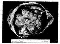
大体:肿瘤直径8~21 cm,8例肿瘤包膜完整,表面光滑,6例肿瘤与周围组织有粘连。切面:肿瘤呈囊性或囊实性,囊内均可见油脂及毛发物。囊内壁可见结节状增厚区或团块状实性区,范围从2~9 cm不等(图1),该区域大部分呈灰白色,两例甲状腺肿恶变者实性区呈淡褐色。 鳞状细胞癌变者部分区域形成乳头或菜花状结构,质地较脆,常伴有出血、坏死。
表1 卵巢成熟性囊性畸胎瘤恶变患者临床病理表现
Table 1 Clinical-pathological feature of ovarian mature cystic teratoma with malignant transformation No
Diagnosis
Age
, 百拇医药
Side
Size
Clinical
stage
Follow up
1
Carcinoid
68
RO
18.0 cm×15.0 cm×6.0 cm
FF
2
, 百拇医药 S.C.C
67
RO
21.0 cm×13.0 cm×11.0 cm
Ⅳ
Alive after 27 months
S.C.C
73
RO
10.0 cm×10.0 cm×7.0 cm
FF
4
, 百拇医药
S.C.C
81
RO
17.0 cm×17.0 cm×12.0 cm
Ⅲ
FF
5
S.C.C
79
RO
8.0 cm×5.0 cm×4.0 cm
Ⅳ
Died after 2 months
, 百拇医药
6
S.C.C
71
RO
8.0 cm×7.0 cm×2.0 cm
Ia
FF
7
S.C.C
79
RO
11.0 cm×11.0 cm×10.0 cm
Ia
, 百拇医药
Died after 5 months
8
S.C.C
82
LO
14.0 cm×10.0 cm×4.0 cm
Ⅰ
Died after 12 months
9
S.C.C
56
RO
, 百拇医药
10.0 cm×5.0 cm×10.0 cm
Ⅲc
Died after 6 months
10
Adeno CA
41
RO
12.0 cm×12.5 cm×9.5 cm
Ⅰa
Alive after 48 months
11
, 百拇医药 S.C.C
30
LO
9.0 cm×10.0 cm×7.0 cm
Ⅱb
Died after 8 months
12
PapCA
43
RO
15.0 cm×8.0 cm×4.0 cm
Ⅰa
, 百拇医药
Alive after 60 months
13
S.C.C
41
LO
10.0 cm×8.0 cm×8.0 cm
Ⅲa
FF
14
S.C.C
53
LO
, http://www.100md.com 9.0 cm×5.5 cm×4.5 cm
Ⅲ
Alive after 6 months
S.C.C, squamous cell carcinoma; PapCA, thyroid papillary carcinoma; Carcinoid, strumous carcinoid; Adeno CA, adenocarcinoma; LO: left ovary; RO, right ovary; FF: fail follow up.
镜下:所有肿瘤均可见成熟性畸胎瘤成分,包括分化好的鳞状上皮、皮肤附属器、脂肪组织、纤维结缔组织等。有11例为鳞状上皮成分恶变,其中9例为高分化的鳞状细胞癌,有7例可以找到良性上皮恶性变的过渡区。另2例鳞状细胞癌组织分化较差,向间质内浸润生长,呈恶性纤维组织细胞瘤样改变。2例成熟性畸胎瘤伴有卵巢甲状腺肿成分,其中1例在甲状腺组织内出现典型的乳头状腺癌结构,另1例在甲状腺肿中出现类癌成分,瘤细胞排列成实性巢和小腺泡状,细胞大小一致,胞浆红染(图2)。免疫组化染色显示NSE及Chromogranin A阳性。1例在成熟性畸胎瘤中出现粘液腺癌成分。
, http://www.100md.com
2.3 HPV检测
对7例鳞状细胞癌病例的宫颈及瘤体组织PCR扩增,PCR产物经40 g.L-1凝胶电泳后分析,除阳性对照外,未发现阳性样本。
图1 卵巢成熟性囊性畸胎瘤鳞状细胞癌变
肿瘤呈囊性改变局部伴有实性区
Figure 1 Squamous cell carcinoma arising in a mature cystic teratoma.
The tumour had a predominantly cystic appearance with focal solid areas.
, 百拇医药
图2 卵巢成熟性囊性畸胎瘤甲状腺肿区中的类癌成分 HE×200
Figure 2 Strumous carcinoid in thyroid tissue
of a mature cystic teratoma HE×200
3 讨论
MCT是最常见的卵巢肿瘤之一,从婴儿期到绝经后的老年妇女均可发病,但以生育期妇女最多见[1~3]。卵巢MCT恶变则较少见,国外报告发生率为2%~4%[2,3],国内报告在2%~5%之间[1]。年龄19~88岁,但多数病例发生在绝经后妇女[1~3]。MCT恶变常发生在单侧卵巢,左、右侧发病率相似。本组病例中13例均为单侧卵巢肿瘤,右侧明显多于左侧(10∶3),1例为双侧卵巢发生的MCT, 其中左侧的肿瘤发生恶变。病理检查,发生恶变的肿瘤体积通常较大,本组病例发生恶变肿瘤的直径在8~21 cm(平均13 cm)。1例双侧卵巢畸胎瘤患者,左侧肿瘤直径为20 cm,发生恶变;而右侧肿瘤直径6 cm, 未发现恶变。恶变常发生在肿瘤的实性区,肉眼检查可见这些区域呈灰白色,质地较脆,常伴有出血坏死。因此,临床上,当病人年龄大、畸胎瘤体积较大时,应注意恶变的发生,术中可制冰冻切片观察以决定进一步的手术方案。在畸胎瘤送检标本中,对实性区应充分取材。发现上述的肉眼改变时,特别是当肿瘤被膜不完整,与周围组织粘连时应高度怀疑恶变的发生。本组病例中,有6例经术中冰冻切片诊断有MCT恶变,1例怀疑有恶变。
, 百拇医药
MCT恶变可以发生在3个胚层,以鳞状细胞癌最为多见,可占75%~88%[1~3,5,6]。本组病例中鳞状细胞癌占78.6% (11/14),其中又以高分化鳞状细胞癌为主(占81.8%, 9/11)。这一型肿瘤细胞分化好,常可见角珠形成,其中7例可找到良性鳞状上皮向恶性转化的过渡区。 2例分化较差的鳞状细胞癌,肿瘤向间质内浸润性生长,部分区域呈恶性纤维组织细胞瘤样改变,免疫组化证实其中的肉瘤样细胞仍具有上皮细胞表达,表明是一种肉瘤样癌的形态,这种形态学改变在MCT恶变中尚未见报告。卵巢畸胎瘤可以完全由甲状腺组织组成,称为卵巢甲状腺肿(struma-ovarii)。甲状腺组织也可作为畸胎瘤的一种成分出现。两者中的甲状腺成分均可发生恶变[7]。本组病例中,2例甲状腺组织均作为畸胎瘤的一种成分出现并发生恶变,其中1例为典型的甲状腺乳头状腺癌,另1例则为较少见的类癌。腺癌在MCT恶变中并不常见[8],本组病例中的1例为粘液腺癌,并可见到癌变区与呼吸上皮有关。
HPV是一种亲上皮的病毒,特别是对鳞状上皮有易感性。已经确定HPV具有转化基因E6和E7[9],这两种基因可以使肿瘤抑制基因p53和pRB丢失正常功能。大约有20种类型的HPV感染与鳞状细胞癌有关的[10]。其中HPV16及HPV18与宫颈上皮内瘤病(cervical intraepithelial neoplasia, CIN)和宫颈鳞状细胞癌有密切关系[10]。然而,有关HPV感染与卵巢肿瘤关系的研究报告还较少。Mai等[11]报告了1例双侧卵巢原发性鳞状细胞癌伴有高度CIN,通过原位杂交法,在外阴、宫颈及左侧卵巢肿瘤中检测出HPV16/18。Mai的结果显示HPV可以通过上行途径感染卵巢。 Sworn等[12]描述了1例卵巢囊肿伴有鳞状上皮内肿瘤,患者同时伴有CIN Ⅲ级。经原位杂交及PCR技术检查,宫颈病变中发现HPV16, 而在卵巢肿瘤中未发现HPV。目前尚不清楚HPV感染与卵巢的MCT恶变、特别是与鳞状细胞癌变有无直接关系。我们对7例MCT鳞状细胞癌变的卵巢肿瘤及宫颈组织应用PCR技术检测 HPV16/18感染情况,结果未发现1例阳性标本。 因此,我们的结果提示HPV感染不是卵巢的MCT鳞状细胞癌变的主要原因。
, 百拇医药
卵巢MCT恶变的机制不清,在我们先前的研究中,发现这一肿瘤有多种染色体畸变[13]。除类癌外,MCT 恶变的预后较差,文献报告5年生存率为13%~30%[1~3]。在本组病例中,3例术中即有周围器官侵犯。在随访的10例病人中,有5人在诊断后1年之内死亡。
(感谢香港大学玛丽医院妇产科实验室刘皙技师在PCR技术上给予的帮助。)
参考文献
1 李 勤,李 静.卵巢.见:陈忠年,杜心谷,刘伯宁.妇产科病理学.上海:上海医科大学出版社,1996. 231-239
2 Talerman A. Germ cell tumours of the ovary. In: Kurman RJ. Blaustein's pathology of the female genital tract. 4th ed. New York: Springer-Verlag, 1994. 849-878
, 百拇医药
3 Nogales FF. Germ cell tumours of the ovary. In: Fox H, ed. Haines & Taylor obstetrical and gynecological pathology. 4th ed. New York: Churchill Livingstone, 1995. 847-871
4 Ip EWK, Collins RJ, Wong RLC, et al. Human papillomavirus and its prognostic significance in invasive carcinoma of the cervix in young patients. Int J Gynecol Path, 1992, 11:158-165
5 Tseng CJ, Chou HH, Huang KG, et al. Squamous cell carcinoma arising in mature cystic teratoma of the ovary. Gynecol Oncol, 1996, 63: 364-370
, 百拇医药
6 Amerigo J, Nogales FF, Fernadez-Sanz J, et al. Squamous cell neoplasm arising from ovarian benign cystic teratoma. Gynecol Oncol, 1997, 8: 277-283
7 Devaney K, Snyder R, Norris HJ, et al. Proliferative and histologically malignant struma ovarii: a clinicopathologic study of 54 cases. Int J Gynecol Pathol, 1993, 12:333-343
8 Malviya VK, Boike GM, Amirikia H, et al. Adenocarcinoma in a mature cystic teratoma. A case report. J Reprod Med, 1989, 34:183-186
, 百拇医药
9 Howley PM. Role of the human papillomavirus in human cancer. Cancer Res, 1991, 51: 5019-5022
10 Yee CL, Krishnan-Hewlett I, Baker CC, et al. Presence and expression of human papillomavirus sequences in human cervical carcinoma cell lines. Am J Pathol, 1985, 119:3261-3266
11 Mai KT, Yazdi HM, Bertrand MA, et al. Bilateral primary ovarian squamous call carcinoma associated with human papilloma virus infection and vulvar and cervical intraepithelial neoplasia. Am J Surg Pathol, 1996, 20: 767-772
, 百拇医药
12 Sworn MJ, Jones H, Letchworth AT, et al. Squamous intraepithelial neoplasia in an ovarian cyst, cervical intraepithelial neoplasia, and human papillomavirus. Hum Pathol, 1995, 26: 344-347
13 Shen DH, Khoo US, Xue WC, et al. Ovarian mature cystic teratoma with malignant transformation an interphase cytogenetic study. Int J Gynecol Pathol, 1998, 17:351-357
(1998-09-16收稿), http://www.100md.com
单位:沈丹华 薛为成 北京医科大学人民医院病理科,北京 100044;颜婉嫦 张雅贤 香港大学玛丽医院
关键词:卵巢肿瘤;病理学;乳头状瘤病毒;人;分离和提纯;畸胎癌;病理
北京医科大学学报990414 摘 要 目的:分析卵巢成熟性囊性畸胎瘤恶变的临床病理学特征及其与人类乳头瘤病毒感染的关系。方法:回顾性研究了14例卵巢成熟性囊性畸胎瘤恶变临床及病理学特征,并且应用PCR技术对其中7例的瘤体组织及宫颈组织进行了人类乳头瘤病毒检测。结果:14例卵巢畸胎瘤恶变病例发病年龄为30~82岁(平均61.7岁),组织学类型包括鳞状细胞癌(11例)、腺癌、甲状腺乳头状腺癌及甲状腺类癌(各1例)。人类乳头瘤病毒检测没有发现阳性病例。结论:卵巢成熟性囊性畸胎瘤恶变是一种少见的病变,发病年龄以绝经后妇女为主,组织学类型以鳞状细胞癌最为多见,未发现HPV感染与其的关系。
, http://www.100md.com
学
中国图书资料分类法分类号 R737.31
Clinicopathological analysis and human papilloma virus status
of ovarian mature cystic teratoma with malignant transformation
SHEN Dan-Hua#, ZHANG Ya-Xian (CHEUNG A N Y), XUE Wei-Cheng, YAN Wan-Chang (NGAN H Y S)
(#Department of Pathology, People's Hospital, Beijing Medical University, Beijing 100044)
, 百拇医药
ABSTRACT Objective: To analyze the clinicopathological feature and status of human papilloma virus (HPV) infection of ovarian mature cystic teratoma with malignant transformation. Methods: The clinical and pathological data of 14 cases ovarian mature cystic teratoma with malignant transformation were analyzed. Detection of HPV in 7 cases of tumors and cervices was performed by PCR technique. Results: The ages of patients ranged from 30 to 82 years (mean, 61.7 years). The tumor size (diameter) ranged from 8 cm to 21 cm (mean, 13 cm). Histologically, there were 11 cases of squamous cell carcinoma (78.6%), one of adenocarcinoma, one of thyroid papillary carcinoma, and one of strumous carcinoid. There was no evidence of HPV infection in the cases studied. Conclusion: Ovarian mature cystic teratoma with malignant teratoma is uncommon and in most cases occurs in postmenopausal women. Squamous cell carcinoma is of the most common histological type. The absence of HPV suggests that there is no association between HPV infection and this tumour.
, http://www.100md.com
MeSH Ovarian neoplasms/pathol Papillomavirus, human/isol Teratocarcinoma/pathol
成熟性囊性畸胎瘤(mature cystic teratoma, MCT)是女性卵巢较常见的肿瘤,占所有卵巢肿瘤的10%~34%[1~3]。本病少数病例可以发生恶性转化,称为成熟性囊性畸胎瘤恶变(MCT with malignant transformation)。虽然畸胎瘤的3个胚层均可发生恶变,但鳞状细胞癌是最常见的组织类型。有许多研究显示人类乳头瘤病毒(human papilloma virus, HPV)感染与生殖系统的鳞状细胞癌有关。我们分析了14例来自北京和香港的卵巢MCT恶变患者的临床及病理学特征,并对其中7例鳞状细胞癌进行HPV检测,以探讨HPV感染与鳞状细胞癌的关系。
1 材料与方法
, 百拇医药 材料来源:收集1983~1998年间14例卵巢MCT恶变病例:其中6例来自北京医科大学人民医院,8例来自香港大学玛丽医院。所有材料均采用100 g.L-1的福尔马林固定,石蜡包埋。
复习及总结临床病历,观察HE切片进行组织学分类[2]。
对7例鳞状细胞癌病例,分别从宫颈及肿瘤组织包埋块中切片提取DNA。使用HPV16、HPV16B、HPV18、HPVL1作为引物,应用PCR扩增技术后,检测样本中的HPV[8]。
2 结果
2.1 临床表现(表1)
14例病人发病年龄为30~82岁(平均61.7岁),其中9例发生在绝经后,1例在更年期,4例为生育期。 病人主要的临床症状是腹胀,腹部包块。2例病人近期消瘦。 12例肿瘤诊断时的临床分期为5例Ⅰ期,1例Ⅱ期,4例Ⅲ期,2例Ⅳ期。13例为单侧卵巢肿物,其中10例位于右侧卵巢,3例位于左侧卵巢;1例为双侧卵巢肿瘤,恶变肿瘤位于左侧卵巢。治疗采用子宫加双附件切除,其中2例进行局部淋巴结清扫。术后化疗。在随访的10例病人中,目前有5例在本病诊断后1年内死亡,另5例存活6个月~4年。
, 百拇医药
2.2 病理所见
大体:肿瘤直径8~21 cm,8例肿瘤包膜完整,表面光滑,6例肿瘤与周围组织有粘连。切面:肿瘤呈囊性或囊实性,囊内均可见油脂及毛发物。囊内壁可见结节状增厚区或团块状实性区,范围从2~9 cm不等(图1),该区域大部分呈灰白色,两例甲状腺肿恶变者实性区呈淡褐色。 鳞状细胞癌变者部分区域形成乳头或菜花状结构,质地较脆,常伴有出血、坏死。
表1 卵巢成熟性囊性畸胎瘤恶变患者临床病理表现
Table 1 Clinical-pathological feature of ovarian mature cystic teratoma with malignant transformation No
Diagnosis
Age
, 百拇医药
Side
Size
Clinical
stage
Follow up
1
Carcinoid
68
RO
18.0 cm×15.0 cm×6.0 cm
FF
2
, 百拇医药 S.C.C
67
RO
21.0 cm×13.0 cm×11.0 cm
Ⅳ
Alive after 27 months
S.C.C
73
RO
10.0 cm×10.0 cm×7.0 cm
FF
4
, 百拇医药
S.C.C
81
RO
17.0 cm×17.0 cm×12.0 cm
Ⅲ
FF
5
S.C.C
79
RO
8.0 cm×5.0 cm×4.0 cm
Ⅳ
Died after 2 months
, 百拇医药
6
S.C.C
71
RO
8.0 cm×7.0 cm×2.0 cm
Ia
FF
7
S.C.C
79
RO
11.0 cm×11.0 cm×10.0 cm
Ia
, 百拇医药
Died after 5 months
8
S.C.C
82
LO
14.0 cm×10.0 cm×4.0 cm
Ⅰ
Died after 12 months
9
S.C.C
56
RO
, 百拇医药
10.0 cm×5.0 cm×10.0 cm
Ⅲc
Died after 6 months
10
Adeno CA
41
RO
12.0 cm×12.5 cm×9.5 cm
Ⅰa
Alive after 48 months
11
, 百拇医药 S.C.C
30
LO
9.0 cm×10.0 cm×7.0 cm
Ⅱb
Died after 8 months
12
PapCA
43
RO
15.0 cm×8.0 cm×4.0 cm
Ⅰa
, 百拇医药
Alive after 60 months
13
S.C.C
41
LO
10.0 cm×8.0 cm×8.0 cm
Ⅲa
FF
14
S.C.C
53
LO
, http://www.100md.com 9.0 cm×5.5 cm×4.5 cm
Ⅲ
Alive after 6 months
S.C.C, squamous cell carcinoma; PapCA, thyroid papillary carcinoma; Carcinoid, strumous carcinoid; Adeno CA, adenocarcinoma; LO: left ovary; RO, right ovary; FF: fail follow up.
镜下:所有肿瘤均可见成熟性畸胎瘤成分,包括分化好的鳞状上皮、皮肤附属器、脂肪组织、纤维结缔组织等。有11例为鳞状上皮成分恶变,其中9例为高分化的鳞状细胞癌,有7例可以找到良性上皮恶性变的过渡区。另2例鳞状细胞癌组织分化较差,向间质内浸润生长,呈恶性纤维组织细胞瘤样改变。2例成熟性畸胎瘤伴有卵巢甲状腺肿成分,其中1例在甲状腺组织内出现典型的乳头状腺癌结构,另1例在甲状腺肿中出现类癌成分,瘤细胞排列成实性巢和小腺泡状,细胞大小一致,胞浆红染(图2)。免疫组化染色显示NSE及Chromogranin A阳性。1例在成熟性畸胎瘤中出现粘液腺癌成分。
, http://www.100md.com
2.3 HPV检测
对7例鳞状细胞癌病例的宫颈及瘤体组织PCR扩增,PCR产物经40 g.L-1凝胶电泳后分析,除阳性对照外,未发现阳性样本。

图1 卵巢成熟性囊性畸胎瘤鳞状细胞癌变
肿瘤呈囊性改变局部伴有实性区
Figure 1 Squamous cell carcinoma arising in a mature cystic teratoma.
The tumour had a predominantly cystic appearance with focal solid areas.

, 百拇医药
图2 卵巢成熟性囊性畸胎瘤甲状腺肿区中的类癌成分 HE×200
Figure 2 Strumous carcinoid in thyroid tissue
of a mature cystic teratoma HE×200
3 讨论
MCT是最常见的卵巢肿瘤之一,从婴儿期到绝经后的老年妇女均可发病,但以生育期妇女最多见[1~3]。卵巢MCT恶变则较少见,国外报告发生率为2%~4%[2,3],国内报告在2%~5%之间[1]。年龄19~88岁,但多数病例发生在绝经后妇女[1~3]。MCT恶变常发生在单侧卵巢,左、右侧发病率相似。本组病例中13例均为单侧卵巢肿瘤,右侧明显多于左侧(10∶3),1例为双侧卵巢发生的MCT, 其中左侧的肿瘤发生恶变。病理检查,发生恶变的肿瘤体积通常较大,本组病例发生恶变肿瘤的直径在8~21 cm(平均13 cm)。1例双侧卵巢畸胎瘤患者,左侧肿瘤直径为20 cm,发生恶变;而右侧肿瘤直径6 cm, 未发现恶变。恶变常发生在肿瘤的实性区,肉眼检查可见这些区域呈灰白色,质地较脆,常伴有出血坏死。因此,临床上,当病人年龄大、畸胎瘤体积较大时,应注意恶变的发生,术中可制冰冻切片观察以决定进一步的手术方案。在畸胎瘤送检标本中,对实性区应充分取材。发现上述的肉眼改变时,特别是当肿瘤被膜不完整,与周围组织粘连时应高度怀疑恶变的发生。本组病例中,有6例经术中冰冻切片诊断有MCT恶变,1例怀疑有恶变。
, 百拇医药
MCT恶变可以发生在3个胚层,以鳞状细胞癌最为多见,可占75%~88%[1~3,5,6]。本组病例中鳞状细胞癌占78.6% (11/14),其中又以高分化鳞状细胞癌为主(占81.8%, 9/11)。这一型肿瘤细胞分化好,常可见角珠形成,其中7例可找到良性鳞状上皮向恶性转化的过渡区。 2例分化较差的鳞状细胞癌,肿瘤向间质内浸润性生长,部分区域呈恶性纤维组织细胞瘤样改变,免疫组化证实其中的肉瘤样细胞仍具有上皮细胞表达,表明是一种肉瘤样癌的形态,这种形态学改变在MCT恶变中尚未见报告。卵巢畸胎瘤可以完全由甲状腺组织组成,称为卵巢甲状腺肿(struma-ovarii)。甲状腺组织也可作为畸胎瘤的一种成分出现。两者中的甲状腺成分均可发生恶变[7]。本组病例中,2例甲状腺组织均作为畸胎瘤的一种成分出现并发生恶变,其中1例为典型的甲状腺乳头状腺癌,另1例则为较少见的类癌。腺癌在MCT恶变中并不常见[8],本组病例中的1例为粘液腺癌,并可见到癌变区与呼吸上皮有关。
HPV是一种亲上皮的病毒,特别是对鳞状上皮有易感性。已经确定HPV具有转化基因E6和E7[9],这两种基因可以使肿瘤抑制基因p53和pRB丢失正常功能。大约有20种类型的HPV感染与鳞状细胞癌有关的[10]。其中HPV16及HPV18与宫颈上皮内瘤病(cervical intraepithelial neoplasia, CIN)和宫颈鳞状细胞癌有密切关系[10]。然而,有关HPV感染与卵巢肿瘤关系的研究报告还较少。Mai等[11]报告了1例双侧卵巢原发性鳞状细胞癌伴有高度CIN,通过原位杂交法,在外阴、宫颈及左侧卵巢肿瘤中检测出HPV16/18。Mai的结果显示HPV可以通过上行途径感染卵巢。 Sworn等[12]描述了1例卵巢囊肿伴有鳞状上皮内肿瘤,患者同时伴有CIN Ⅲ级。经原位杂交及PCR技术检查,宫颈病变中发现HPV16, 而在卵巢肿瘤中未发现HPV。目前尚不清楚HPV感染与卵巢的MCT恶变、特别是与鳞状细胞癌变有无直接关系。我们对7例MCT鳞状细胞癌变的卵巢肿瘤及宫颈组织应用PCR技术检测 HPV16/18感染情况,结果未发现1例阳性标本。 因此,我们的结果提示HPV感染不是卵巢的MCT鳞状细胞癌变的主要原因。
, 百拇医药
卵巢MCT恶变的机制不清,在我们先前的研究中,发现这一肿瘤有多种染色体畸变[13]。除类癌外,MCT 恶变的预后较差,文献报告5年生存率为13%~30%[1~3]。在本组病例中,3例术中即有周围器官侵犯。在随访的10例病人中,有5人在诊断后1年之内死亡。
(感谢香港大学玛丽医院妇产科实验室刘皙技师在PCR技术上给予的帮助。)
参考文献
1 李 勤,李 静.卵巢.见:陈忠年,杜心谷,刘伯宁.妇产科病理学.上海:上海医科大学出版社,1996. 231-239
2 Talerman A. Germ cell tumours of the ovary. In: Kurman RJ. Blaustein's pathology of the female genital tract. 4th ed. New York: Springer-Verlag, 1994. 849-878
, 百拇医药
3 Nogales FF. Germ cell tumours of the ovary. In: Fox H, ed. Haines & Taylor obstetrical and gynecological pathology. 4th ed. New York: Churchill Livingstone, 1995. 847-871
4 Ip EWK, Collins RJ, Wong RLC, et al. Human papillomavirus and its prognostic significance in invasive carcinoma of the cervix in young patients. Int J Gynecol Path, 1992, 11:158-165
5 Tseng CJ, Chou HH, Huang KG, et al. Squamous cell carcinoma arising in mature cystic teratoma of the ovary. Gynecol Oncol, 1996, 63: 364-370
, 百拇医药
6 Amerigo J, Nogales FF, Fernadez-Sanz J, et al. Squamous cell neoplasm arising from ovarian benign cystic teratoma. Gynecol Oncol, 1997, 8: 277-283
7 Devaney K, Snyder R, Norris HJ, et al. Proliferative and histologically malignant struma ovarii: a clinicopathologic study of 54 cases. Int J Gynecol Pathol, 1993, 12:333-343
8 Malviya VK, Boike GM, Amirikia H, et al. Adenocarcinoma in a mature cystic teratoma. A case report. J Reprod Med, 1989, 34:183-186
, 百拇医药
9 Howley PM. Role of the human papillomavirus in human cancer. Cancer Res, 1991, 51: 5019-5022
10 Yee CL, Krishnan-Hewlett I, Baker CC, et al. Presence and expression of human papillomavirus sequences in human cervical carcinoma cell lines. Am J Pathol, 1985, 119:3261-3266
11 Mai KT, Yazdi HM, Bertrand MA, et al. Bilateral primary ovarian squamous call carcinoma associated with human papilloma virus infection and vulvar and cervical intraepithelial neoplasia. Am J Surg Pathol, 1996, 20: 767-772
, 百拇医药
12 Sworn MJ, Jones H, Letchworth AT, et al. Squamous intraepithelial neoplasia in an ovarian cyst, cervical intraepithelial neoplasia, and human papillomavirus. Hum Pathol, 1995, 26: 344-347
13 Shen DH, Khoo US, Xue WC, et al. Ovarian mature cystic teratoma with malignant transformation an interphase cytogenetic study. Int J Gynecol Pathol, 1998, 17:351-357
(1998-09-16收稿), http://www.100md.com