神经激肽B受体在猫三叉神经脊束核尾侧亚核和脊髓内的分布*
作者:吕葆真 丁玉强 王丹 张淼丽
单位:吕葆真 丁玉强 王丹 张淼丽(第四军医大学解剖学教研室,梁琚脑研究中心,西安 710032)
关键词:神经激肽B受体;三叉神经尾侧亚核;脊髓;免疫组织化学;猫
解剖学报990302
【摘要】 目的 了解神经激肽B受体(NK3受体)与伤害性信息的传递和调制的可能关系,调查NK3受体在三叉神经尾侧亚核和脊髓内的分布状况。 方法 免疫组织化学技术。 结果 NK3受体样免疫反应产物在三叉神经尾侧亚核和脊髓内的分布基本一致。致密的免疫反应产物分布于Ⅱ内层(inner part),而Ⅰ层和Ⅱ外层(outer part)染色浅淡。NK3受体阳性神经元胞体主要见于三叉神经尾侧亚核和脊髓的Ⅰ层和Ⅱ层,少量分布于脊髓后角深层。 结论 提示三叉神经尾侧亚核和脊髓内的NK3受体可能与初级伤害性信息的传递和调控有密切关系。
, 百拇医药
DISTRIBUTION OF NEUROKININ B RECEPTOR (NK3) IN THE CAUDAL
SPINAL TRIGEMINAL NUCLEUS AND SPINAL CORD OF THE CAT Baozhen, Ding Yuqiang△, Wang Dan, Zhang Miaoli
(Department of Anatomy and K.K.Leung Brain Research Center,The Fourth Military Medical University, Xi'an)
【Abstract】 Objectives In order to study possible relationship between neurokinin B receptor(NK3) and transmission and modulation of noxious sensory information, we investigated the distribution of NK3 receptor in the caudal spinal trigeminal nucleus and spinal cord of the cat. Method Immunohistochemical staining was used. Results The distribution pattern of NK3 receptor-like immunoreactivity (-LI) in the spinal cord was similar to that of the caudal spinal trigeminal nucleus. Intense NK3 receptor-LI was observed in the inner part of lamina Ⅱ of the caudal spinal trigeminal nucleus and spinal cord; Weak NK3 receptor-LI was seen in lamina Ⅰ and outer part of lamina Ⅱ. Neuronal cell bodies with NK3 receptor-LI were mainly found in laminae Ⅰ and Ⅱ of the caudal spinal trigeminal nucleus and spinal dorsal horn, and a few were distributed in the deeper layers of the spinal dorsal horn. Conclusion The results suggest that NK3 receptor might be involved in the transmission and modulation of primary noxious information in the caudal spinal trigeminal nucleus and spinal cord of the cat.
, 百拇医药
【Key words】 Neurokinin B receptor; Caudal spinal trigeminal nucleus; Spinal cord; Immunohistochemistry; Cat
速激肽家族主要由P物质、K物质和神经激肽B(NKB,亦称nenromedin K)组成,它们广泛分布于神经系统内,并具有多种重要的生理功能[1]。3种速激肽的生理作用是通过各自的受体介导实现的,即P物质(SP)受体(NK1)、K物质受体(NK2)和NKB受体(NK3)[2]。迄今的研究表明,神经系统内NKB及其受体NK3参与调节机体多种内脏功能活动,同时也与伤害性信息的传递和调制有密切关系。脊髓和延髓后角(三叉神经尾侧亚核)是神经系统接受外周伤害性刺激的重要区域,在形态学上弄清NK3受体在脊髓和延髓后角的分布状况,是阐明其与伤害性信息的传递和调制的关系不可缺少的。因此,本研究采用免疫组织化学技术观察了NK3受体在猫三叉神经尾侧亚核和脊髓内的分布状况。
, 百拇医药
材料和方法
实验共用成年家猫3只,在腹腔内注射戊巴比妥钠(100mg/kg)的深麻醉状态下,开胸经升主动脉插管,先用500 ml生理盐水冲净血液,然后用1 000 ml含4%的多聚甲醛和0.5%苦味酸的0.1 mol/L磷酸缓冲液(PB, pH7.4)灌注固定1h,灌毕取出脑干和脊髓及半月节和背根节(L7、S1),并在上述固定液内后固定2h(4℃),然后置于含有30%蔗糖的0.1 mol/L PB内(pH7.4)过夜。冰冻冠状切片(厚30μm)。所选取的脊髓节段为C5、T6、L2、L7和S2 5个节段。切片经0.01 mol/L磷酸盐缓冲液(PBS,pH 7.4)漂洗后,依此进入:(1)兔抗NK3受体(0.5mg/L,关于此抗体的制备和鉴定请参照文献[3]);(2)生物素标记的羊抗兔IgG(Vector, 1:200);(3)ABC复合物(Vector,1:200)。其中,1抗和2抗用含0.9%驴血清和0.3%的Triton X-100的0.01mol/L的PBS(pH 7.4)稀释,ABC复合物用0.3%TritonX-100的0.001mol/L的PBS(pH 7.4)稀释。最后切片在含0.05%DAB和0.003%的H2O2的0.05 mol/L Tris-HCl缓冲液(pH 7.6)内呈色20~30min。
, http://www.100md.com
对照实验在省略1抗或用正常兔血清代替1抗的情况下进行,结果为阴性。
结果和讨论
免疫组织化学染色结果显示,NK3受体样免疫反应产物在三叉神经尾侧亚核和脊髓内的分布基本一致。
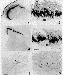
在三叉神经尾侧亚核内,致密的免疫反应产物分布于Ⅱ内层,而Ⅰ层和Ⅱ外层染色浅淡(图1)。在高倍镜下观察发现,三叉神经尾侧亚核1~Ⅲ层内分布有一定数量的NK3受体阳性神经元,绝大部分为圆形或三角形的小细胞(<10μm),少量中等大小的神经元仅见于Ⅰ层内(图2)。此外,在Ⅲ层的外侧份还可见到由Ⅱ层延伸而来的阳性突起(图2)。在三叉神经尾侧亚核的其他部分,包括三叉脊束内均未见有阳性反应产物出现。半月节内的神经节细胞为阴性。
在所观察的脊髓节段内(C5、T6、L2、L7和S2),NK3受体样免疫反应产物分布均与三叉神经尾侧亚核相似。致密的免疫反应产物出现于脊髓后角Ⅱ内层,其次为Ⅰ层和Ⅱ外层(图3,4)。但阳性神经元的分布与尾侧亚核不同,一是Ⅰ~Ⅲ层内阳性神经元数量少于尾侧亚核(图4),二是在Ⅴ~Ⅶ层和中央管周围灰质(Ⅹ层)内均发现有少量散在分布的中等大小的阳性神经元。这些神经元着色浅淡,它们的突起亦呈现NK3受体样免疫反应,酷似Golgi染色外观(图5,6)。此外,在前角、胸腰节段的中间带外侧核和中央管周围灰质内还发现有一定数量的免疫反应纤维,但前角和中间带外侧核内未见有免疫反应胞体。后根节、背外侧束、后索、侧索和前索内均未发现有免疫反应产物出现。
, 百拇医药
大鼠脑内NK3受体分布的研究表明,三叉神经尾侧亚核和脊髓内NK3受体样免疫反应产物亦主要分布于Ⅱ层内[3],且证实阳性产物定位于神经元的胞体和树突内,轴突和胶质细胞为阴性[4]。本研究发现NK3受体主要分布于猫三叉神经尾侧亚核和脊髓Ⅱ内层,这种分布上的差别可能是由于动物种属差异造成的。大鼠[3]和猫的三叉神经半月节和后根节内均无NK3受体阳性细胞,且在脊髓白质包括背外侧束及三叉神经脊束内也未观察到有NK3受体免疫反应纤维。因此,推测三叉神经尾侧亚核和脊髓尤其是浅层内NK3受体阳性神经元可能为中间神经元,而三叉神经尾侧亚核和脊髓Ⅱ内层的免疫反应结构可能主要为浅层内阳性神经元的树突。这有待免疫电镜的研究提供进一步的证据。
大量关于速激肽及其受体的研究表明,它们在伤害性信息传递中起着重要的作用。NK3受体在大鼠和猫均集中分布于三叉神经脊束核和脊髓后角Ⅱ层,而Ⅱ层主要是由大量接受伤害性信息的中间神经元组成。因此,三叉神经尾侧亚核和脊髓浅层内的NK3受体可能与伤害性信息的传递和调制有密切关系。最近的研究提示,NKB/NK3受体可能与SP/NK1受体的作用相反,具有抗伤害性感觉的作用[5]。
, http://www.100md.com
本文照片的制作得到了原悦萍同志的协助,谨致谢意。
图1,2 示NK3受体样免疫反应产物在猫三叉神经脊束核尾侧亚核内的分布。致密的反应产物位于Ⅱ内层(Ⅱi),其次为Ⅰ层、Ⅱ外层(Ⅱ0)和Ⅲ层。Ⅰ~Ⅲ层内还可见有NK3受体阳性细胞。图2为图1中尾侧亚核浅层的放大像。标尺示320μm(图1)和30μm(图2)
图3,4 示NK3受体样免疫反应产物在猫颈髓后角内的分布。致密的反应产物位于Ⅱ内层(Ⅱi),其次为Ⅰ层、Ⅱ外层(Ⅱ0)和Ⅲ层。图4为图3后角浅层的放大像。标尺示320μm(图3)和30μm(图4)
图5,6 示腰髓(L7)后角V层和颈髓(C5)中央管周围灰质内NK3受体阳性细胞(箭头)。标尺示20μm
, http://www.100md.com
Fig.1,2 NK3 receptor-like immunoreactivity in the caudal spinal trigeminal nucleus of the cat. Intense NK3 receptor-LI is located in the inner part of lamina Ⅱ, and weak NK3 receptor-LI is seen in lamina Ⅰ, outer part of lamina Ⅱ, and lamina Ⅲ. NK3 receptor-positive neurons are distributed in laminae Ⅰ-Ⅲ. The superficial layers of the caudal spinal trigeminal nucleus in Fig.1 is enlarged in Fig.2. Bar=320 μm in Fig.1 and 30 μm in Fig.2
, http://www.100md.com
Fig.3,4 NK3 receptor-like immunoreactivity in the fifth cervical spinal horn of the cat. Intense NK3 receptor-LI is located in the inner part of lamina Ⅱ, and weak NK3 receptor-LI is seen in lamina Ⅰ, outer part of lamina Ⅱ, and lamina Ⅲ. The superficial layer of the dorsal horn in Fig.3 is enlarged in Fig.4. Bar=320 μm in Fig.3 and 30 μm in Fig.4.
Fig.5,6 NK3 receptor-positive neurons (arrows) in lamina Ⅴ of the seventh lumbar spinal cord and the region around the central canal of the fifth cervical spinal cord. Bar=20 μm
, 百拇医药
* 国家自然科学基金资助课题(No.39600045)
△ Department of Anatomy and K.K. Leung Brain Research Center, The Fourth Military Medical University, Xi'an 710032, China
参考文献
[1]Otsuka M, Yoshioka K.Neurotransmitter functions of mammalian tachykinins. Physiol Rev, 1993, 73(2):229
[2]Nakanishi S.Mammalian tachykinin receptors. Annu Rev Neurosci, 1991, 14(1):123
, http://www.100md.com
[3]Ding YQ, Shigemoto R, Takada M,et al. Localization of the neuromedin K receptor (NK3) in the central nervous system of the rat. J Comp Neurol, 1996, 364(1):290
[4]Seybold VS, Grkovic I, Portbury AL, et al. Relationship of NK3 receptor-immunoreactivity to subpopulations of neurons in rat spinal cord. J Comp Neurol, 1997, 381(2):439
[5]McCarson KE, Krause JE. The formalin-induced expression of tachykinin peptide and neurokinin receptor messenger RNAs in rat sensory ganglia and spinal cord is modulated by opiate preadministration. Neuroscience, 1995, 64(2):729
收稿1998-03 修回1998-08, http://www.100md.com
单位:吕葆真 丁玉强 王丹 张淼丽(第四军医大学解剖学教研室,梁琚脑研究中心,西安 710032)
关键词:神经激肽B受体;三叉神经尾侧亚核;脊髓;免疫组织化学;猫
解剖学报990302
【摘要】 目的 了解神经激肽B受体(NK3受体)与伤害性信息的传递和调制的可能关系,调查NK3受体在三叉神经尾侧亚核和脊髓内的分布状况。 方法 免疫组织化学技术。 结果 NK3受体样免疫反应产物在三叉神经尾侧亚核和脊髓内的分布基本一致。致密的免疫反应产物分布于Ⅱ内层(inner part),而Ⅰ层和Ⅱ外层(outer part)染色浅淡。NK3受体阳性神经元胞体主要见于三叉神经尾侧亚核和脊髓的Ⅰ层和Ⅱ层,少量分布于脊髓后角深层。 结论 提示三叉神经尾侧亚核和脊髓内的NK3受体可能与初级伤害性信息的传递和调控有密切关系。
, 百拇医药
DISTRIBUTION OF NEUROKININ B RECEPTOR (NK3) IN THE CAUDAL
SPINAL TRIGEMINAL NUCLEUS AND SPINAL CORD OF THE CAT
 Baozhen, Ding Yuqiang△, Wang Dan, Zhang Miaoli
(Department of Anatomy and K.K.Leung Brain Research Center,The Fourth Military Medical University, Xi'an)
【Abstract】 Objectives In order to study possible relationship between neurokinin B receptor(NK3) and transmission and modulation of noxious sensory information, we investigated the distribution of NK3 receptor in the caudal spinal trigeminal nucleus and spinal cord of the cat. Method Immunohistochemical staining was used. Results The distribution pattern of NK3 receptor-like immunoreactivity (-LI) in the spinal cord was similar to that of the caudal spinal trigeminal nucleus. Intense NK3 receptor-LI was observed in the inner part of lamina Ⅱ of the caudal spinal trigeminal nucleus and spinal cord; Weak NK3 receptor-LI was seen in lamina Ⅰ and outer part of lamina Ⅱ. Neuronal cell bodies with NK3 receptor-LI were mainly found in laminae Ⅰ and Ⅱ of the caudal spinal trigeminal nucleus and spinal dorsal horn, and a few were distributed in the deeper layers of the spinal dorsal horn. Conclusion The results suggest that NK3 receptor might be involved in the transmission and modulation of primary noxious information in the caudal spinal trigeminal nucleus and spinal cord of the cat.
, 百拇医药
【Key words】 Neurokinin B receptor; Caudal spinal trigeminal nucleus; Spinal cord; Immunohistochemistry; Cat
速激肽家族主要由P物质、K物质和神经激肽B(NKB,亦称nenromedin K)组成,它们广泛分布于神经系统内,并具有多种重要的生理功能[1]。3种速激肽的生理作用是通过各自的受体介导实现的,即P物质(SP)受体(NK1)、K物质受体(NK2)和NKB受体(NK3)[2]。迄今的研究表明,神经系统内NKB及其受体NK3参与调节机体多种内脏功能活动,同时也与伤害性信息的传递和调制有密切关系。脊髓和延髓后角(三叉神经尾侧亚核)是神经系统接受外周伤害性刺激的重要区域,在形态学上弄清NK3受体在脊髓和延髓后角的分布状况,是阐明其与伤害性信息的传递和调制的关系不可缺少的。因此,本研究采用免疫组织化学技术观察了NK3受体在猫三叉神经尾侧亚核和脊髓内的分布状况。
, 百拇医药
材料和方法
实验共用成年家猫3只,在腹腔内注射戊巴比妥钠(100mg/kg)的深麻醉状态下,开胸经升主动脉插管,先用500 ml生理盐水冲净血液,然后用1 000 ml含4%的多聚甲醛和0.5%苦味酸的0.1 mol/L磷酸缓冲液(PB, pH7.4)灌注固定1h,灌毕取出脑干和脊髓及半月节和背根节(L7、S1),并在上述固定液内后固定2h(4℃),然后置于含有30%蔗糖的0.1 mol/L PB内(pH7.4)过夜。冰冻冠状切片(厚30μm)。所选取的脊髓节段为C5、T6、L2、L7和S2 5个节段。切片经0.01 mol/L磷酸盐缓冲液(PBS,pH 7.4)漂洗后,依此进入:(1)兔抗NK3受体(0.5mg/L,关于此抗体的制备和鉴定请参照文献[3]);(2)生物素标记的羊抗兔IgG(Vector, 1:200);(3)ABC复合物(Vector,1:200)。其中,1抗和2抗用含0.9%驴血清和0.3%的Triton X-100的0.01mol/L的PBS(pH 7.4)稀释,ABC复合物用0.3%TritonX-100的0.001mol/L的PBS(pH 7.4)稀释。最后切片在含0.05%DAB和0.003%的H2O2的0.05 mol/L Tris-HCl缓冲液(pH 7.6)内呈色20~30min。
, http://www.100md.com
对照实验在省略1抗或用正常兔血清代替1抗的情况下进行,结果为阴性。
结果和讨论
免疫组织化学染色结果显示,NK3受体样免疫反应产物在三叉神经尾侧亚核和脊髓内的分布基本一致。
在三叉神经尾侧亚核内,致密的免疫反应产物分布于Ⅱ内层,而Ⅰ层和Ⅱ外层染色浅淡(图1)。在高倍镜下观察发现,三叉神经尾侧亚核1~Ⅲ层内分布有一定数量的NK3受体阳性神经元,绝大部分为圆形或三角形的小细胞(<10μm),少量中等大小的神经元仅见于Ⅰ层内(图2)。此外,在Ⅲ层的外侧份还可见到由Ⅱ层延伸而来的阳性突起(图2)。在三叉神经尾侧亚核的其他部分,包括三叉脊束内均未见有阳性反应产物出现。半月节内的神经节细胞为阴性。
在所观察的脊髓节段内(C5、T6、L2、L7和S2),NK3受体样免疫反应产物分布均与三叉神经尾侧亚核相似。致密的免疫反应产物出现于脊髓后角Ⅱ内层,其次为Ⅰ层和Ⅱ外层(图3,4)。但阳性神经元的分布与尾侧亚核不同,一是Ⅰ~Ⅲ层内阳性神经元数量少于尾侧亚核(图4),二是在Ⅴ~Ⅶ层和中央管周围灰质(Ⅹ层)内均发现有少量散在分布的中等大小的阳性神经元。这些神经元着色浅淡,它们的突起亦呈现NK3受体样免疫反应,酷似Golgi染色外观(图5,6)。此外,在前角、胸腰节段的中间带外侧核和中央管周围灰质内还发现有一定数量的免疫反应纤维,但前角和中间带外侧核内未见有免疫反应胞体。后根节、背外侧束、后索、侧索和前索内均未发现有免疫反应产物出现。
, 百拇医药
大鼠脑内NK3受体分布的研究表明,三叉神经尾侧亚核和脊髓内NK3受体样免疫反应产物亦主要分布于Ⅱ层内[3],且证实阳性产物定位于神经元的胞体和树突内,轴突和胶质细胞为阴性[4]。本研究发现NK3受体主要分布于猫三叉神经尾侧亚核和脊髓Ⅱ内层,这种分布上的差别可能是由于动物种属差异造成的。大鼠[3]和猫的三叉神经半月节和后根节内均无NK3受体阳性细胞,且在脊髓白质包括背外侧束及三叉神经脊束内也未观察到有NK3受体免疫反应纤维。因此,推测三叉神经尾侧亚核和脊髓尤其是浅层内NK3受体阳性神经元可能为中间神经元,而三叉神经尾侧亚核和脊髓Ⅱ内层的免疫反应结构可能主要为浅层内阳性神经元的树突。这有待免疫电镜的研究提供进一步的证据。
大量关于速激肽及其受体的研究表明,它们在伤害性信息传递中起着重要的作用。NK3受体在大鼠和猫均集中分布于三叉神经脊束核和脊髓后角Ⅱ层,而Ⅱ层主要是由大量接受伤害性信息的中间神经元组成。因此,三叉神经尾侧亚核和脊髓浅层内的NK3受体可能与伤害性信息的传递和调制有密切关系。最近的研究提示,NKB/NK3受体可能与SP/NK1受体的作用相反,具有抗伤害性感觉的作用[5]。
, http://www.100md.com
本文照片的制作得到了原悦萍同志的协助,谨致谢意。

图1,2 示NK3受体样免疫反应产物在猫三叉神经脊束核尾侧亚核内的分布。致密的反应产物位于Ⅱ内层(Ⅱi),其次为Ⅰ层、Ⅱ外层(Ⅱ0)和Ⅲ层。Ⅰ~Ⅲ层内还可见有NK3受体阳性细胞。图2为图1中尾侧亚核浅层的放大像。标尺示320μm(图1)和30μm(图2)
图3,4 示NK3受体样免疫反应产物在猫颈髓后角内的分布。致密的反应产物位于Ⅱ内层(Ⅱi),其次为Ⅰ层、Ⅱ外层(Ⅱ0)和Ⅲ层。图4为图3后角浅层的放大像。标尺示320μm(图3)和30μm(图4)
图5,6 示腰髓(L7)后角V层和颈髓(C5)中央管周围灰质内NK3受体阳性细胞(箭头)。标尺示20μm
, http://www.100md.com
Fig.1,2 NK3 receptor-like immunoreactivity in the caudal spinal trigeminal nucleus of the cat. Intense NK3 receptor-LI is located in the inner part of lamina Ⅱ, and weak NK3 receptor-LI is seen in lamina Ⅰ, outer part of lamina Ⅱ, and lamina Ⅲ. NK3 receptor-positive neurons are distributed in laminae Ⅰ-Ⅲ. The superficial layers of the caudal spinal trigeminal nucleus in Fig.1 is enlarged in Fig.2. Bar=320 μm in Fig.1 and 30 μm in Fig.2
, http://www.100md.com
Fig.3,4 NK3 receptor-like immunoreactivity in the fifth cervical spinal horn of the cat. Intense NK3 receptor-LI is located in the inner part of lamina Ⅱ, and weak NK3 receptor-LI is seen in lamina Ⅰ, outer part of lamina Ⅱ, and lamina Ⅲ. The superficial layer of the dorsal horn in Fig.3 is enlarged in Fig.4. Bar=320 μm in Fig.3 and 30 μm in Fig.4.
Fig.5,6 NK3 receptor-positive neurons (arrows) in lamina Ⅴ of the seventh lumbar spinal cord and the region around the central canal of the fifth cervical spinal cord. Bar=20 μm
, 百拇医药
* 国家自然科学基金资助课题(No.39600045)
△ Department of Anatomy and K.K. Leung Brain Research Center, The Fourth Military Medical University, Xi'an 710032, China
参考文献
[1]Otsuka M, Yoshioka K.Neurotransmitter functions of mammalian tachykinins. Physiol Rev, 1993, 73(2):229
[2]Nakanishi S.Mammalian tachykinin receptors. Annu Rev Neurosci, 1991, 14(1):123
, http://www.100md.com
[3]Ding YQ, Shigemoto R, Takada M,et al. Localization of the neuromedin K receptor (NK3) in the central nervous system of the rat. J Comp Neurol, 1996, 364(1):290
[4]Seybold VS, Grkovic I, Portbury AL, et al. Relationship of NK3 receptor-immunoreactivity to subpopulations of neurons in rat spinal cord. J Comp Neurol, 1997, 381(2):439
[5]McCarson KE, Krause JE. The formalin-induced expression of tachykinin peptide and neurokinin receptor messenger RNAs in rat sensory ganglia and spinal cord is modulated by opiate preadministration. Neuroscience, 1995, 64(2):729
收稿1998-03 修回1998-08, http://www.100md.com