大鼠Meynert基底核注入海人藻酸后c-fos原癌基因蛋白在脑纹状体边缘区及边缘系统内的表达*
作者:包新民 舒斯云 李胜修 张运周 周光斗 吴文龙
单位:包新民 舒斯云 李胜修 张运周 周光斗 吴文龙(第一军医大学珠江医院神经科学研究所,广州,510282)
关键词:纹状体边缘区;c-fos;Meynert氏基底核
大鼠Meynert基底核注入海人藻酸后c 摘要:为研究Meynert基底核与纹状体边缘区以及脑内其他跟学习记忆有关的边缘系统结构间的关系,将海人藻酸注射于大鼠Meynert基底核后,观察c-fos原癌基因蛋白在脑内的表达。海人藻酸注射4h后,脑内只有少量c-fos阳性表达;海人藻酸注射8h后,脑内c-fos原癌基因蛋白表达增至最高峰。在海马、齿状回、中央杏仁核、纹状体、边缘区中有大量密集的c-fos阳性表达。在终纹床核、丘脑网状核、外侧隔核、丘脑下部视上核、室旁核、前下托及嗅内皮质等处可见较多c-fos阳性表达。在伏隔核、视前区、丘脑底核、黑质外侧部等部位可见少量c-fos阳性表达。海人藻酸注射Meynert基底核16h后,除海马、齿状回、中央杏仁核、纹状体及边缘区中有较多表达外,其余部位阳性标记基本消失。但脑腹侧的梨状前皮质、嗅结节、杏仁皮质核及前嗅核等部位出现大量的c-fos阳性表达。大脑皮质也可见较多c-fos阳性表达。海人藻酸注射48h后,脑内各部位的c-fos阳性反应均消失。本研究结果提示Meynert基底核和纹状体边缘区以及跟脑内其它与学习记忆有关的边缘系统结构间有密切的功能联系。
, http://www.100md.com
中图分类号: R741
Expression of oncogene c-fos protein in the marginal division and limbic system following kainic acid injection into the basal nucleus of Meynert in rat Bao Xinmin, Shu Siyun, Li Shengxiu, Zhang Yunzhou, Zhou Guangdou, Wu Wenlong
Institute for Neuroscience, Zhujiang Hospital, First Military Medical University, Guangzhou, 510282
Abstract: To investigate the relationship between marginal division of striatum and other structures related to learning and memory function of the brain, 0.1% kainic acid was stereotaxically injected into the Meynert’s basal nucleus, and the expression of oncogene c-fos protein in the brain was observed by using the immunohistochemistry. 4h later, there were few c-fos-positive cells in the brain; 8h later, expression of c-fos was increased at the peak. Many dark stained c-fos-positive neurons were found in marginal division of the striatum, hippocampus, dentate gyrus, central amygdaloid nucleus, striatum, reticular nucleus of the thalamus, the bed nucleus of the stria terminals, thalamic reticular nucleus, lateral septal nucleus, supraoptic nucleus, paraventricular nucleus of hypothalamus, presubiculum and entorhinal cortex. A few c-fos-positive reactions were found in the accumbens, preoptic area, subthalamic nucleus, lateral part of substantia nigra. 16h later, the positive expression disappeared, except in the hippocampus, dentate gyrus, central amygdaloid nucleus, striatum, and marginal division of striatum. However, numerous c-fos-positive cells were observed in the prepiriform cortex, olfactory tubercle, cortical amygdaloid nucleus, anterior olfactory nucleus and cortex. 48h later, all the c-fos reaction in the brain disappeared. These results suggest that the function connections exist between the Meynert’s basal nucleus or marginal division and limbic structures which are concerned with learning and memory function. Therefore, it is assumed that the marginal division is related to the function of learning and memory of the brain.
, 百拇医药
Key words: marginal division of striatum; c-fos; Meynert’s basal nucleus
Meynert基底核发出的胆碱能纤维投射到大脑皮层和海马等广大区域,损伤后影响动物的学习记忆功能[1,2]。纹状体边缘区是我们在纹状体中发现的一个新亚区[3]。它的细胞形态、化学递质、突触特征等均不同于纹状体的其它部分[4~7]。迷宫试验证明纹状体边缘区的功能跟学习、记忆有关[8]。我们曾观察到[9,10]纹状体边缘区和Meynert基底核、黑质外侧部及其背外侧区有纤维联系。并用c-fos表达法观察到[11]纹状体边缘区和Meynert基底核以及脑内与学习、记忆功能有关的海马、基底前脑、皮质等边缘系统结构之间有功能联系。
为进一步研究纹状体边缘区和Meynert基底核及脑边缘系统之间的关系,本文用海人藻酸注射大鼠Meynert基底核后,观察c-fos原癌基因蛋白在脑内的表达,尤其是在纹状体边缘区以及其它和学习、记忆功能有关的结构如海马及杏仁核等边缘系统中表达的情况。
, 百拇医药
1 材料和方法
实验动物为SD雄性大鼠,共38只,体质量180~250g,购自第一军医大学实验动物中心。动物房温度维持在18~26℃,光暗周期为12h。实验前在上述条件下喂养(颗粒型大鼠饲料+自来水)两周,以适应饲养环境。动物随机分为6组:
1.1 Meynert基底核注射组 将20只腹腔内注射10%水合氯醛麻醉(4ml/kgb.w.)后的大鼠固定在立体定位仪上,参照包新民等[12]的图谱,取Meynert基底核的座标为AP=B-1.4,ML=±3.2,H=6.8。以尖端外径为50mm的微玻管向Meynert基底核内缓慢注射0.1%海人藻酸0.2ml,留针15min。动物分别存活2(2只)、4(3只)、8(5只)、16(5只)、24(3只)、48h(2只)后灌注取材。在麻醉下经升主动脉先灌注生理盐水100ml,再灌注含4%多聚甲醛的0.1mol/L低温磷酸缓冲液500ml。取脑后放入含30%蔗糖的磷酸缓液内置冰箱中过夜。然后将脑进行冰冻冠状或矢状切片,片厚40mm,每隔2张取1张切片,进行以下免疫组化步骤。(1)0.3%Triton X-10030min,室温。(2)5%正常羊血清20min,室温。(3)兔抗c-fos血清(INCSTAR公司,1:1000),37℃孵育30min后在4℃冰箱中孵育48h。(4)生物素标记的羊抗兔IgG(Vector,1:200),4~5h,室温。(5)SP(Zymed,1:200)4~5h,室温。以上各步骤间均以0.01mol/L磷酸盐缓冲液洗涤4×15min。然后用0.1mol/L醋酸缓冲液(pH6.0)浸泡15min,进行GDN呈色反应。切片经裱片、干燥、透明、封片后在Olympus AH3显微镜下观察、照相。
, 百拇医药
1.2 纹状体注射对照组 大鼠5只,按第一组的方法将0.1%海人藻酸注射于纹状体的中部。动物分别存活4(2只)、8(2只)或16h(1只)后同上进行灌注取材及免疫组化过程。
1.3 苍白球注射对照组 大鼠5只,按第一组的方法将0.1%海人藻酸注射于苍白球部位。动物分别存活4(2只)、8(2只)或16h(1只)后同上进行灌注取材及免疫组化过程。
1.4 载体注射对照组 用生理盐水0.2ml代替海人藻酸注射于Meynert基底核,存活4(2只)或8h(2只)后进行上述实验。
1.5 麻醉对照组 大鼠经腹腔内注射10%水合氯醛(4ml/kg)进行麻醉后,分别存活4(2只)或8h(2只)后再进行上述实验。
1.6 免疫组化对照组 在海人藻酸注射于Meynert基底核组中,取一套切片以正常羊血清代替抗c-fos血清进行上述免疫组化反应。
, 百拇医药
2 结果
2.1 海人藻酸注射于Meynert基底核后2h,脑组织切片上未见c-fos阳性反应。动物存活4h后,在大鼠脑的中央杏仁核及海马等处有少量c-fos阳性表达。动物存活8h后,脑内c-fos原癌基因蛋白表达增至最高峰。在纹状体及纹状体边缘区中有大量密集的c-fos阳性表达,在边缘区中的表达非常清楚,呈明显的条带状(图1~3)。在杏仁中央核,海马结构的CA1和齿状回中也有大量密集的c-fos阳性表达(图4)。在嗅内皮质、前下托、终纹床核、外侧隔核、丘脑网状核、丘脑下部视上核和室旁核中可见较多c-fos阳性表达(图4,5)。另外,在视前区、伏隔核、苍白球、丘脑底核、黑质外侧部等处可见少量c-fos阳性表达。海人藻酸注射Meynert基底核后16h,除海马、齿状回、中央杏仁核、纹状体及边缘区中有较多表达外,其余部位阳性标记基本消失。但脑腹侧的梨状前皮质、嗅结节、杏仁皮质核及前嗅核等部位出现大量的c-fos阳性表达(图6~8)。大脑皮质也可见较多c-fos阳性表达。海人藻酸注射后48h,脑内各部位的c-fos阳性反应均消失。
, 百拇医药
2.2 对照实验结果 海人藻酸注射于纹状体中部时,动物存活4~6h后,在梨状前皮质和大脑皮质有较多c-fos阳性表达,在纹状体、丘脑中线核等处有少量c-fos阳性表达。海人藻酸注射于苍白球后存活4~6h的动物,在梨状前皮质和大脑皮质中可见较多c-fos阳性表达。抗体空白对照实验组及麻醉对照实验组的动物,在各存活期的各脑区中均未见c-fos原癌基因蛋白阳性表达。载体注射对照组的动物在梨状前皮质中有很少量c-fos阳性表达,但和实验组有显著性差异(P<0.01)。
图1 海人藻酸注射Meynert基底核后8h,在纹状体边缘区中出现大量密集的c-fos阳性表达
Fig.1 Strong positive expression of c-fos in the marginal division of the striatum, 8 h after kainic acid injection into the Meynert's basal nucleus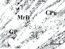
, 百拇医药
图2 图1的放大,示边缘区中的c-fos阳性细胞
Fig.2 Magnification of figure 1, showing the c-fos-positive cells in the MrD
图3 图2的放大,MrD边缘区
Fig.3 Magnification of figure 2, MrD marginal division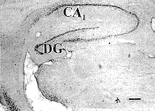
, 百拇医药
图4 海人藻酸注入Meynert基底核后8h,在海马、齿状回及前下托中有大量密集的c-fos阳性表达
Fig.4 Strong positive expression of c-fos in the hippocampus, dentate gyrus, and presubiculum, 8 h after kainic acid injection into the Meynert's basal nucleus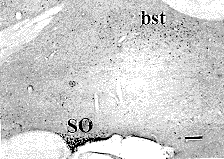
图5 海人藻酸注入Meynert基底核后8h,在视上核中有大量c-fos阳性表达,终纹床核中有少量c-fos阳性表达
Fig. 5 After 8 h of kainic acid injection into the Meynert's basal nucleus, stronger positive expression of c-fos in the supraoptic nucleus and weaker positive expression of c-fos in the bed nucleus of the stria terminals
, 百拇医药
图6 海人藻酸注入Meynert基底核后16h,除海马及齿状回中有较多c-fos阳性表达外,在下托及杏仁皮质核中出现大量c-fos阳性表达
Fig. 6 After 16 h of kainic acid injection into the Meynert's basal nucleus, strong positive expression in the subiculum and cortical nucleus of amygdala, strong c-fos positive expression in the hippocampus and dentate gyrus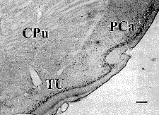
图7 海人藻酸注入Meynert基底核后16h,在梨状前皮质前部及嗅结节中出现大量c-fos阳性表达
, 百拇医药
Fig. 7 After 16 h of kainic acid injection into the Meynert's basal nucleus, strong positive expression of c-fos in the anterior part of prepiriform cortex and olfactory tubercle
图8 海人藻酸注入Meynert基底核后16h,在前嗅核及嗅结节中出现大量c-fos阳性表达
Fig.8 After 16 h of kainic acid injection into the Meynert's basal nucleus, strong positive expression of c-fos in the anterior olfactory nucleus and olfactory tubercle
, 百拇医药
Fig.1~8 Bright field microphotographs of rat brain, sagital section, bar=50 m m. Aco: Anterior cortical nucleus of amygdala; aod: Anterior olfactory nucleus, dorsal part; Aov: Anterior olfactory nucleus, ventral part; bst: Bed nucleus of stria terminals; CA1: Field CA1 of the Ammon's horn; Ce: central amygdaloid nucleus; CPu: caudate putamen; DG: Dentate gyrus; GP: globus pallidus; ic: Internal capsule; MrD: Marginal division; PCa: anterior part of prepiriform cortex; S: Subiculum; SO: Supraoptic nucleus; TU: Olfactory tubercle.
, 百拇医药
3 讨论
c-fos原癌基因是一种快速、短暂表达的基因,当中枢神经元受到物理或化学等各种刺激后,激活受体及第二信使,诱导c-fos原癌基因在胞浆中转录成mRNA,并合成c-fos原癌基因蛋白,c-fos原癌基因蛋白进入胞核中与目的基因结合,激活目的基因的表达而对刺激作出反应[13]。由于c-fos原癌基因蛋白仅在和受刺激的神经元有功能联系的神经元中表达,并且用免疫组化或原位杂交等方法容易显示。因此,c-fos原癌基因蛋白表达法是一种较好的研究神经系统功能和结构相结合的方法。最近,Sugimoto等[14]将海人藻酸注射于纹状体吻侧后,在苍白球、脚内核、黑质、丘脑底核等处见到c-fos原癌基因蛋白的表达。而Ciani等[15]观察到将海人藻酸注射于纹状体时,在嗅皮质、杏仁核、海马、扣带回、丘脑中线核等多处见到c-fos原癌基因蛋白的表达。他们观察到的表达部位的差异可能和注射于纹状体的部位及注射的剂量不同等有关。我们曾将微量的海人藻酸注射纹状体边缘区后,在Meynert基底核及边缘系统许多部位见到c-fos原癌基因蛋白的表达[11]。本研究将海人藻酸注射于Meynert基底核后,观察到在纹状体边缘区内有大量密集的c-fos原癌基因蛋白的阳性表达,呈明显的条带状分布。提示纹状体边缘区和Meynert基底核之间有交互的功能联系。另外,Meynert基底核注射海人藻酸后,在海马及杏仁核等处有大量密集的c-fos原癌基因蛋白的阳性表达,提示Meynert基底核和边缘系统之间也有密切的功能联系。因此,推测纹状体边缘区可直接或通过Meynert基底核和边缘系统联系。从对照组的结果来看,注射Meynert基底核和注射纹状体及苍白球后,梨状前皮质和大脑皮质均有大量c-fos原癌基因蛋白的阳性表达,但纹状体和苍白球注射后早期即出现表达,而Meynert基底核注射后16h才明显表达,可能是通过纹状体或苍白球间接表达的。
, 百拇医药
基底前脑尤其是Meynert基底核损伤后可造成动物学习记忆功能障碍[1,2]。我们用迷宫试验已证明边缘区的功能跟学习、记忆有关[8]。本研究观察到当海人藻酸注射于Meynert基底核后,在纹状体边缘区中见到大量密集的c-fos原癌基因蛋白的阳性表达。在与学习、记忆有关的结构如海马、基底前脑、杏仁核等处均有c-fos原癌基因蛋白的阳性表达。再次证明边缘区与脑内跟学习、记忆功能有关的结构间有密切的功能联系,但边缘区在脑的学习、记忆功能中的地位以及与其他学习、记忆有关结构间的关系尚待进一步研究。
*国家自然科学基金(39770550)和广东省科学基金(950554)资助
作者简介:包新民,男1933年出生;1956年毕业于中国医科大学;教授;电话:85143636
**舒斯云(项目负责人)
, 百拇医药
参考文献
1 Mayo W, Kharouby M, Le Moal M et al. Memory disturbances following ibotenic acid injections in the nucleus basalis magnocellularis of the rat. Brain Res, 1988, 455 (2):213
2 Miyamoto M, Shintani M, Nagaoka A et al. Lesioning of the rat basal forebrain leads to memory impairments in passive and active avoidance tasks. Brain Res, 1985, 328 (1):97
3 Shu SY, Penny GR, Peterson GM. The “Marginal Division”: a new sub-division in the neostriatum of the rat. J Chem Neuroanat, 1988, 1 (3):147
, http://www.100md.com
4 Shu SY, McGinty JF, Peterson GM. High density of zinc-containing and dynorphin B- and substance P-immunoreactive terminals in the marginal division of the rat striatum. Brain Res Bull, 1990, 24 (2):2201
5 包新民,舒斯云. 大鼠纹状体边缘区内P物质、脑啡肽和胆囊收缩素免疫阳性反应的分布. 神经解剖学杂志,1997, 13 (2):107
6 包新民,舒斯云. 大鼠纹状体边缘区内神经降压肽和生长抑素样免疫阳性反应的分布. 中国组织化学和细胞化学杂志, 1997, 6(1):1
7 Shu SY, Bao X M, Peterson GM. Ultrastructure of the marginal division in the rat striatum. Chin Med J, 1992, 105(2):102
, 百拇医药
8 李胜修,舒斯云,包新民等. 海人藻酸损毁纹状体边缘区后对大鼠学习和记忆功能影响的研究. 神经解剖学杂志, 1996, 12 (1):37
9 包新民,舒斯云,李胜修等. 大鼠纹状状体边缘区的传入投入- WGA-HRP法研究. 神经解剖学杂志, 1993, 9 (1):93
10 舒斯云,包新民,李胜修等. 大鼠纹状体边缘区传入投射的免疫组化研究. 解剖学杂志, 1993, 16 (1):509
11 包新民,舒斯云,张运周. 海人藻酸注射大鼠纹状体边缘区后c-fos原癌基因在脑内的表达. 神经解剖学杂志,1997, 13 (4):343
12 包新民,舒斯云. 大鼠脑立体定位图谱. 北京:人民卫生出版社, 1991. 120
13 Bullit E. Expression of c-fos-like protein as a marker for neuronal act- ivity following noxious stimulation in the rat. J Comp Neurol, 1990, 296 (4):517
, 百拇医药
14 Sugimoto T, Sato K, Houtani T et al. On the expression of Fos-like protein in the subthalamic nucleus and basal ganglia output systems following kainic acid injections into the rodent striatum. Neurosci Lett, 1993, 152 (1-2):25
15 Ciani E, Guarnieri T. Contestabible A: Fos protein induction, neuropathology, and pharmacological protection after excitotoxic brain insult. Exp Brain Res, 1994, 98 (3):421
(收稿日期:1999-03-18), 百拇医药