马肉孢子虫的包囊超微结构及其实验感染终末宿主的研究
作者:胡俊杰 左仰贤
单位:胡俊杰(云南大学档案学系,昆明 650091);左仰贤(云南大学生物系,昆明 650091)
关键词:马肉孢子虫;自然感染率;超微结构;实验感染
寄生虫与医学昆虫学报000204摘要 昆明地区马肉孢子虫包囊的自然感染率为78%。光镜下。包囊呈梭形,具有锥状和棍棒状的突起。透射电镜下突起内的微管在顶端松散排列,向下延伸集结成束,斜伸入基质层,有的和母细胞的膜相连。用含有包囊的马肉实验感染2只犬、2只猫,感染后犬粪中发现孢子囊和卵囊。潜伏期为13~14天。剖检,2只犬的小肠固有层中均查到孢子囊和卵囊。猫粪中一直未检到孢子囊和卵囊,剖检亦未发现。表明马肉孢子虫的终末宿主是犬,不是猫。我们认为马体内的肉孢子虫仅有一种,鉴定为柏氏肉孢子虫(Sarcocystis bertrami)。本文是国内马肉孢子虫的首次报告。
, 百拇医药
PREVALENCE,CYST WALL ULTRASTRUCTURES AND EXPERIMENTAL
DIFINITIVE HOST INFECTIONS OF SARCOCYSTIS FROM HORSES
Hu Junjie
(Department of Biology,Yunnan University,Kumming,650091)
Zuo Yangxian
(Department of Biology,Yunnan University,Kumming,650091)
Abstract The prevalence of sarcosporidiosis in horses from Kunming was 76.7%.Light microscropy re-vealed that the intramuscular cysis were shuttle-shaped.The cyst wall was formed by cone-or clubshaped protrusions.Electvo-microscopy showed that in the core of the protrusion there were bundles of microtubules which penetrated diagonally into the ground substance along the longitudinal axis of the protrusion and sometimes reached the interior border of the ground substance.Two dogs that had been fed on musculature containins Sarcocysts of naturally infected horse shed sporulated oocysts and sporocysts in feces 13 to 14 day. later In histologic sections of canine small intestien,the oocysts and sporocysts were located in the lamina porpia near the tips of the vfilli.Two cats fed on similar musculature containing sarcocysts did nof shed sporulated oocysts and sporocysts,ond there were no oocysts and sporocysts in cat small intestine.under histo logical examinations. So the dog is the final host of Sarcocystis of the horse.Based on this study and the literature,we suqqest that only one species named Sarcocystis bertrami exists in horses in Kunming.
, 百拇医药
Key words Horse Sarcocystis Prevalence Ultrastructure Experimental infectivity
在欧洲、亚洲、美洲等地均有关于马肉孢子虫的报道。迄今共报道了有3种寄生于马肌肉中的肉孢子虫:柏氏肉孢子虫(Sarcocystis bertrami)、马犬肉孢子虫(Sarcocystis equicanis)、法氏肉孢子虫(Sarcocysitis fayeri)。关于各个种之间的关系及种名的有效性存在着不同的观点。此外,近年来还在北美马的脑和脊索神经细胞中发现神经肉孢子虫(Sarcocystis neurora),其终末宿主尚不明。我国尚无马肉孢子虫的研究报道。因此,本研究在理论上和实践上具有一定意义。
1 材料与方法
1.1 自然感染率调查及光镜观察包囊
, 百拇医药 分部位剪取昆明市郊屠宰的马肉,带回实验室。剔除脂肪和结缔组织,顺肌纤维方向将肌肉剪成铅笔芯粗细的小条,有规则地置放在载玻片,每张玻片可放5~10条,用另一张载玻片盖上,缓缓轻压,使肌肉变薄变扁,低倍镜下检查肉孢子虫包囊。查到包囊后,记下位置,取去上面一张载玻片,将包囊剥离出来,移入另一张载玻片上,滴加0.85%的生理盐水,加盖玻片,高倍镜或油镜下观察包囊形态、测量和初步鉴定。刺破包囊可观察和测量溢出的缓殖子和母细胞。统计马各部位肌肉的肉孢子虫自然感染率。
1.2 透射电镜材料制备
选包囊较多的一小块马肌肉,以2.5%戊二醛(0.1mol/L pH=7.3的磷酸缓冲液配制)预固定2h,1%锇酸后固定2h,经脱水包埋后作超薄切片,以醋酸双氧铀和柠檬酸铅染色,自然干燥,透射电镜观察。
1.3 实验感染
1.实验动物 犬(Canis familiaris)4只,猫(Felis catus)4只:购自昆明市郊农贸市场,刚断奶,约40日龄。犬和猫实验感染前均经连续5次粪检(每天检查一次),检查肉孢子虫孢子囊和卵囊,证实没有自然感染的肉孢子虫卵囊和孢子囊。
, 百拇医药
2.实验感染 从自然感染的马肉中分离包囊,包含有大小两型包囊,经口感染2只犬和2只猫(约200包囊/只),另2只犬、2只猫留作对照。自感染后第5天起,每天收集犬、猫粪便,以37%ZnSO4漂浮法涂片镜检。15~20天后剖杀动物,取其小肠粘膜固有层涂片镜检。
2 结果
2.1 马肉孢子虫自然感染率调查结果
本研究共对50头马进行了肌肉检查,在39头马的肌肉中发现了肉孢子虫包囊,自然感染率为78%。食道肌、颈部肌、颌肌、舌肌、背肌、肋间肌、膈肌、前腿肌、后腿肌、臀肌、尾肌等12个部位肌肉中均检查到肉孢子虫包囊。常见的寄生部位是食道肌(60.0%)、前腿肌(41.6%)和后腿肌(38.4%)。心肌不被寄生。
2.2 包囊在光镜下的观察结果
, 百拇医药
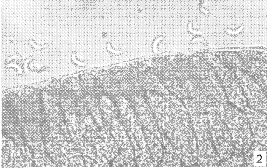
马体内发现两型包囊,两型包囊均呈乳白色,长梭形,具有倾斜指状突起,突起末端伸入肌细胞质。Ⅰ型包囊较小,大小为1 175~7 375μm×25~125μm,平均2 541μm×71.1 μm(n=30)。突起长2.5~5.3μm(含基质层厚度),平均长3.7μm(n=20)。缓殖子香蕉形,大小为14.5~17.0μm×2.1~4.5μm,平均15.0μm×3.9μm(n=20)(图Ⅱ-1)。Ⅱ型包囊较大,大小为1 250~8 325μm×100~325μm,平均4 907μm×241μm(n=24)。突起稍短,突起长1.8~3.0μm(含基质层厚度),平均2.4μm(n=18)。缓殖子香蕉形,大小为15.1~20.5μm×3.8~5.5μm,平均16.3μm×4.2μm(n=20)(图版Ⅱ-2)。


, 百拇医药
1 自然感染的马的柏氏肉孢子虫(Sarcocystis bertrami)Ⅰ型包囊,包囊壁上有长指状突起(×500);
2 自然感染的马的柏氏肉孢子虫(Sarcocystis bertrami)Ⅱ型包囊,包囊壁上有短指状突起(×600);
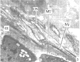
3 马体内具有长指状突起的Ⅰ型包囊的纵切面.突起内有微管(MT)和嗜锇颗粒(OS),微管在突起顶部和中部比较分散,在基部和基质层(GS)集结成束,斜伸入基质层,有时甚至达到基质层的内壁(×13000);
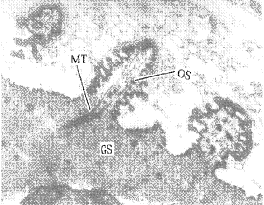
4 马体内具有短指状突起的Ⅰ型包囊的纵切面.突起内有微管(MT)和嗜锇颗粒(OS),微管在突起顶部和中部比较分散,在基部和基质层(GS)集结成束,斜伸入基质层,有时甚至达到基质层的内壁(×26000);
5 感染的柏氏肉孢子虫(Sarcocystis bertrami)14天后,犬粪中的肉孢子虫孢子囊(×1200);
, 百拇医药
6 感染的柏氏肉孢子虫(Sarcocystis bertrami)14天后,犬小肠固有层中的肉孢子虫虫卵囊(×1200);
1 Sarcocysts from naturally infected horse.Sarcocystis bertrami cyst surrounded by a cyst wall with long protrusions(×1500)2.Sarcocystis bertrami cyst surrounded by a cyst wall with short protrusins (×600).3 TEM micrographs of the cyst wall with long protrusions.Longitudinal section through villar protrusion with large osmiophilic granules(OS) and microtubules(MT)in the core.The bundle of microtubules which penetrated diagonally into the ground substance (GS)along the longitudinal axis of the villar protrusion and sometimes reached the interior border of the ground substance was generally loosely organized within the middle and distal parts of the villar protrusions,and tightly packed within the proximal part of the protrusions and the ground substance of the cyst wall (×1300).4 TEM micrographs of the cyst wall with long protrusions.Note osmiophilic granules(OS),microtubules(MT) adn round substance(GS)(×26000).5 Sarcocystis bertrami Sporocysts from the feces of a dong 14 days after feeding horse muscles(×1200).6 Sarcocystis bertrami Sporuiated ocysts from the small intestine of a dog 14 days after feeding horse muscles(×1200).
, 百拇医药
2.3 包囊超微结构
2.3.1 马体内两型肉孢子虫包囊突起在透射电镜下均为指状,突起顶端稍尖。突起表面凹陷较深,基质层表面凹陷较浅。突起内有微管,微管在突起顶部和中部比较分散,在基部和基质层内集结成束,斜伸入基质层,有时到达基质层内壁。微管在集结成束前呈波形,集结成束后呈直线形。突起内沿微管具有平行排列的嗜锇颗粒(图版Ⅱ-3,4))。Ⅰ型包囊突起大小为1.7~4.4μm×0.2~0.4μm,平均2.6μm×0.3μm(不含基质层厚度)。基质层厚度为0.3~0.8μm,平均0.6μm。Ⅱ型包囊突起大小为0.9~2.4μm×0.3~1.0μm,平均1.4μm×0.6μm(不含基质层厚度)。基质层厚度为0.3~1.60μm,平均0.9μm。包囊突起之间的间隔,Ⅰ型包囊约为0.1μm,Ⅱ型包囊约为0.3μm。
2.3.2 马的两型肉孢子虫包囊在超微结构上Ⅰ型包囊突起长而细,Ⅱ型包囊突起短而宽,和光镜的观察结果一致。两型包囊的超微结构都属于Ⅱ型(Dubey et al.,1989),即包囊突起呈指状,突起内有微管,微管在突起中部和基部集结成束,斜伸入基质层,有时到达基质层内壁。
, 百拇医药
2.3.3 实验感染 实验感染的2只犬先后在粪便中排出孢子囊和卵囊(图版Ⅱ-5),其潜伏期为13~14天,剖检,小肠粘膜固有层中也查到孢子囊和卵囊(图版Ⅱ-6)。孢子囊椭圆形,含有4个子孢子和颗粒状的孢子囊余体。孢子囊的大小为10.4~14.5μm×7.5~10.0μm,平均12.4μm×8.6μm(n=27)。实验感染的2只猫,粪检未发现孢子囊和卵囊,剖检,小肠粘膜固有层也未查到孢子囊和卵囊。对照的2只犬和2只猫粪检及剖检均未发现孢子囊和卵囊。
3 讨论
Fayer(1983)、Cawthom(1990)、Trab-Darbatz(1994)分别报道了马的肉孢子虫病,病马常表现贫血、发热、脱毛、厌食、表情淡漠、步态僵直等症状,严重时可导致死亡。昆明地区马的肉孢子虫自然感染率为78%,高于Gunn和Fraher(1992)报道爱尔兰的46%,Kirmse(1986)报道摩洛哥的46.2%、Gobel和Rommel(1980)报道德国的23%,Erber和Geisel(1981)报道德国的15.5%,低于Rommel和Geisel(1975)在德国报道的100%。
, http://www.100md.com
马肉中的肉孢子虫包囊首先被Siedamgrotzky(1872)发现,原始描述是包囊长3~4mm,最长达到12mm,宽0.3mm,有纤毛状突起,长2μm。Doflein(1901)在德国马肉中发现肉孢子虫包囊,并把它命名为柏氏肉孢子虫(Sarcocystis bertrami),包囊长9~10mm,具棒状突起。Rommel和Geisel(1975)在德国马肉中发现肉孢子虫包囊,包囊壁薄、光滑,并通过实验感染证明了犬为其终末宿主,把它命名为马犬肉孢子虫(Sarcocystis equicanis)。Dubey(1977)在美国发现马的肉孢子虫,根据包囊形态、孢子囊大小和潜伏期与马犬肉孢子虫的不同,把它命名为法氏肉孢子虫(Sarcocystis fayeri);认为马犬肉孢子虫是柏氏肉孢子虫的同种异名。值得注意的是Rommel和Geisel、Dubey等人当时都没有进行超微结构的研究。直到1980年,Gobel在德国做了马犬肉孢子虫的超微结构观察,同年,Tinling在美国也做了法氏肉孢子虫的超微结构观察。结果显示,这两种肉孢子虫包囊的超微结构都属于11型(Dubey et al,1989)。我们进行的超微结构研究亦证实了昆明地区马的肉孢子虫包囊的超微结构属于11型(Dubey et al.,1989)。文献中关于马的肉孢子虫研究结果简要摘录见表1。
, 百拇医药
表1 关于马的肉孢子虫研究结果的摘录
Tab.1 Selected properties of sarcocystis in the horse 作者
Authors
种名
Designation
中间
宿主
Interme
diate
host
包囊大小
, 百拇医药
(μm)
Cyst dimensions
包囊壁形态
Cyst wall morphology
突起内嗜
锇颗粒
Large
granules
(TEM)
缓殖子大小
(μm)
Bradyzoite
, 百拇医药
dimensions
终末
宿主
Final
host
光镜下形态
LM
电镜下类型
TEM type
Siedagrotzky
Psrosperimial
马
, 百拇医药 3 000~4 000×300
壁上有纤毛状突起,—
—
16×5
—
(1872)
Tubes
长2μm
Doflein
柏氏肉孢子虫
马
9 000~10 000
棒状突起
, http://www.100md.com
—
—
6
—
(1901)
(S.bertrami)
(长)
(宽)
Rommel et al.
马犬肉孢子虫
马
350
壁薄,光滑
, 百拇医药
—
—
—
犬
(1977)
(S.equicanis)
(长)
Dubey et al.
法氏肉孢子虫
马
900×70
具突起,长1~2μm
—
, 百拇医药
—
15~20
犬
(1977)
(S.fayen)
×2~3
Gobel et al.
马犬肉孢子虫
马
350
具突起,长2~3μm
11
无
, http://www.100md.com
—
犬
(1980)
(S.equicanis)
(长)
Tinling et al.
法氏肉孢子虫
马
990×136
具突起,长1~3μm
11
有
120~16.1×
, 百拇医药
—
(1980)
(S.fayen)
2.8~3.8
Cawthom et al.
法氏肉孢子虫
马
50~500×50~150
具突起,1.3~1.8μm
11
有
8.8~12.3×
, 百拇医药
—
(1990)
(S.fayen)
×0.2~0.4μm
1.9~2.7
Odening et al.
Sarcocystis.sp
马
3 840×272
具突起,长5~6μm
11
?
, 百拇医药
16.6×3.4
—
(1995)
本次实验
柏氏肉孢子虫
马
3 724×156
具突起,长3.5μm
11
有
14.5~20.5×
犬
This study
, http://www.100md.com
(S.bertrami)
2.1~5.5
“-”表示未做观察 国外报道的实验感染结果和本次实验的感染结果证明马的肉孢子虫的终末宿主是犬而不是猫。文献上关于马的肉孢子虫孢子囊的的大小和潜伏期的摘录见表2。
迄今为止,报道的寄生在马肌肉内的肉孢子虫共有3种:柏氏肉孢子虫(S.bertrami)、马犬肉孢子虫(S.equicanis)、法氏肉孢子虫(S.fayeri),这3个种都是以犬为终末宿主,根据包囊大小、包囊壁的结构、孢子囊的大小和潜伏期的长短,分为不同的种,但存在着不同的观点:(1)Dubey(1977)、Levine(1986)认为有2个种,柏氏肉孢子虫(S.bertrami)和法氏肉孢子虫(S.fayeri);马犬肉孢子虫(S.equicanis)是柏氏肉孢子虫(S.bertrami)的同种异名。(2)Hihaidy(1982)、Tenter(1995)认为只有1 个种,柏氏肉孢子虫(S.bertrami)。(3)Odening(1995)提出了两种分类的可能性:a. 仅为一个种,柏氏肉孢子虫(S.bertrami)=马犬肉孢子虫(S.equicanis)=法氏肉孢子虫(S.fayeri);b. 根据包囊突起内是否有嗜锇颗粒,分成2个种:柏氏肉孢子虫(S.bertrami)=马犬肉孢子虫(S.equicanis),突起内无嗜锇颗粒。法氏肉孢子虫(S.fayeri),突起内有嗜锇颗粒。
, http://www.100md.com
表2 关于马肉孢子虫孢子囊大小、潜伏期的摘录
Tab.2 Summaries of Sporocysts dimenstons and prepatent perlods about Sarcocystts in the horse 作者
Authous
种名
Designation
中间宿主
Intermediate
host
终末宿主
Final
, http://www.100md.com
host
潜伏期(天)
Prepatent
Period(days)
孢子囊大小(μm)
Sporocysts dimensions
(μm)
Rommel et al.
马犬肉孢子虫
马
犬
8
, 百拇医药
15~16.3×8.18~11.3
(1975)
(S.equiaonis)
Dubey et al.
法氏肉孢子虫
马
犬
11~14
11~11.3×7.0~8.3
(1977)
(S.fayeri)
Erber et al.
, 百拇医药
Sarcocystis sp
马
犬
11~17
12~14.4×9.3~10.5
(1981)
Fayeri
法氏肉孢子虫
马
犬
10
13.2~15.0×9.5~11.3
, 百拇医药
(1983)
(S.fayeri)
Matuschka
Sarcocystis.sp
马
犬
9~10
12.2~13.8×9.2×9.9
(1983)
Matuschka
马肉孢子虫
马
, http://www.100md.com
犬
10
12.2~13.8×9.2~9.9
(1986)
(S.bertrami)
Juyal et al.
马犬肉孢子虫
马
犬
7
17~20.4×10.2~13.6
(1993)
, http://www.100md.com
(S.equiaonis)
本次实验
马肉孢子虫
马
犬
13~14
10.4~14.5×7.5~10.0
This study
(S.bertrcami)
产生马肉孢子虫分类比较混乱的原因,可能主要是由于定种时描述的不充分所造成的误解和研究者的测量方法不同,也由于没有考虑在不同生长阶段包囊所呈现的不同特征。根据Matuschka等1986年的实验感染中间宿主马的报道。柏氏肉孢子虫(S.bertrami)包囊在马体内1年后为2mm长,持续生长超过34个月。3年后其包囊大小为2.5~9.0mm。Fayer(1983)做了中间宿主马的实验感染,发现包囊突起随着年龄的增长而缩短,感染后127天发现某些包囊突起比较长,某些包囊突起比较短,感染后157天和184天检查,仅发现短突起的包囊。本研究在马肌肉内发现的Ⅱ型包囊,其包囊大小和突起长短的变化可能亦与包囊的年龄有关。
, http://www.100md.com
根据我们的研究和文献中的报道,我们认为马肌肉内的肉孢子虫仅有一个种,即柏氏肉孢子虫(S.bertrami)。Ⅰ型包囊和Ⅱ型包囊是S.bertrami不同发育时期的包囊。马犬肉孢子虫(S.equicanis)和法氏肉孢子虫(S.fayeri)是其同种异名。
Yamada(1993)在日本马肉中发现并报道的Sarcocystis.sp包囊突起呈毛发状,超微结构类型属于Ⅶ型(Dubey et al.,1989),包囊突起底部呈钟形,中部指状,顶端细线状,突起从中部开始弯曲90°,几乎和包囊表面平行。这样的Ⅶ型包囊仅在日本报道过1例。昆明地区马体内未发现此型包囊。
国家自然科学基金资助项目
参考文献
1,左仰贤.1992.球虫学——畜禽和人体的球虫与球虫病.天津:天津科学技术出版社.
, 百拇医药
2,Cawthom, R.J.,M.Clark, R.Hudson et al.1990.Histological and ultrastructural appearance of severe Sarcocystis fayeri infection in a malnourished horse.J.Vet.Diagn.Invest.2:342~345.
3,Dubey,J.P.,R.H. Streitel, P.C. Stromberg et al. 1977.Sarcocystis fayeri sp.n.from the horse. J.Parastitol.63:443~447.
4,Dubey, J.P.,C.A.Speer, R Fayer 1989.Sarcocystosis of animals and man.CRC Press.Edwards,G.T. 1984.Prevalence of equine Sarcocystis in British horses and a comparison of two detection methods.Vet.Res.115:265~267.
, http://www.100md.com
5,Erber,M. and O. Geisel 1981.Vorkommen und Entwicklung von 2 Sarkosporidienarten des Pferden.Z.Parasitenk.65:283~291.
6,Fayer,R. and J.P.Dubey 1982.Sarcocystis:Development of Sarcocystis fayeri in the horse. J.Parasitol.68:856~860.
7,Fayer, R., C. Hounsel, R.C.Giles, 1983.Chromic illness in a Sarcocystis-infected pony.Vet.Rec.113:216~217.
8,Gobel, E. and M. Rommel 1980, Light and electron microscropic study on cysts of Sarcocystis eqicanis in the oesophageal musculature of horses. Berl.Munch.Wschr.93:41~47.
, 百拇医药
9,Gunn,H.M and J.P.Fraher 1992.Incidence of Sarcocystis in skeletal muscles of horses.Vet.Parastiol.42(1~2):33~40.
10,Juyal,P.D., I. S. Kalra and P.P.Gunta 1993.A preliminary report on the development of Sarcocystis equicanis cysts from infected equine muscullature in a dog.Indian.Vet.Med.J.17:70~71.
11,Kirmse,P. 1986.Sarcosporidiosis in equines of Morocco.Br.Vet.J.142:70~72.
12,Levine,N.D. 1986.The taxonomy of Sarcocystis (Protoza, Apicomplexa) species.J.Parasitol.72(3):372~382.
, 百拇医药
13,Matuschka,F.R., T.Schnieder, A. Daugschies et al. 1986.Cyclic transmission of Sarcocystis bertrami Doflein.1901 by the dog to the horse.Protistologica 22:231~233.
14,Odening K., H.H. Wesemeier, G.Walter et al.1995. Ultrastructure of Sarcocystis frome Equids.Acta.Parasitologica.40(1):12~20.
15,Seneviratna, P., G.Edward and L.Degiusti 1975. Frequency of Sarcocystis spp in Detroit,Metropolitan area,Michigan. Am.J.Vet.Res.36(3):337~339.
, 百拇医药
16,Tenter,A.M.1995.Currentresearch on Sarcocystis sp of domestic animals.Inter. J.Parasitol.25(11):1311~1330.
17,Tinling,S.P., G.H.Cardinet, L.L. Blythe, et al.1980.Alight and electron microscropic study of sarcocysts in a horse.J.Parasitol.66:458~465.
18,Yamada,M., H.Yukawa, M.Kenmotsu et al.1993.Studies on the morphology of Sarcocystis in thoroughbred horses in Japan.J.Protozool.Res.3:14~19.
收稿日期:1999-09-06, 百拇医药
单位:胡俊杰(云南大学档案学系,昆明 650091);左仰贤(云南大学生物系,昆明 650091)
关键词:马肉孢子虫;自然感染率;超微结构;实验感染
寄生虫与医学昆虫学报000204摘要 昆明地区马肉孢子虫包囊的自然感染率为78%。光镜下。包囊呈梭形,具有锥状和棍棒状的突起。透射电镜下突起内的微管在顶端松散排列,向下延伸集结成束,斜伸入基质层,有的和母细胞的膜相连。用含有包囊的马肉实验感染2只犬、2只猫,感染后犬粪中发现孢子囊和卵囊。潜伏期为13~14天。剖检,2只犬的小肠固有层中均查到孢子囊和卵囊。猫粪中一直未检到孢子囊和卵囊,剖检亦未发现。表明马肉孢子虫的终末宿主是犬,不是猫。我们认为马体内的肉孢子虫仅有一种,鉴定为柏氏肉孢子虫(Sarcocystis bertrami)。本文是国内马肉孢子虫的首次报告。
, 百拇医药
PREVALENCE,CYST WALL ULTRASTRUCTURES AND EXPERIMENTAL
DIFINITIVE HOST INFECTIONS OF SARCOCYSTIS FROM HORSES
Hu Junjie
(Department of Biology,Yunnan University,Kumming,650091)
Zuo Yangxian
(Department of Biology,Yunnan University,Kumming,650091)
Abstract The prevalence of sarcosporidiosis in horses from Kunming was 76.7%.Light microscropy re-vealed that the intramuscular cysis were shuttle-shaped.The cyst wall was formed by cone-or clubshaped protrusions.Electvo-microscopy showed that in the core of the protrusion there were bundles of microtubules which penetrated diagonally into the ground substance along the longitudinal axis of the protrusion and sometimes reached the interior border of the ground substance.Two dogs that had been fed on musculature containins Sarcocysts of naturally infected horse shed sporulated oocysts and sporocysts in feces 13 to 14 day. later In histologic sections of canine small intestien,the oocysts and sporocysts were located in the lamina porpia near the tips of the vfilli.Two cats fed on similar musculature containing sarcocysts did nof shed sporulated oocysts and sporocysts,ond there were no oocysts and sporocysts in cat small intestine.under histo logical examinations. So the dog is the final host of Sarcocystis of the horse.Based on this study and the literature,we suqqest that only one species named Sarcocystis bertrami exists in horses in Kunming.
, 百拇医药
Key words Horse Sarcocystis Prevalence Ultrastructure Experimental infectivity
在欧洲、亚洲、美洲等地均有关于马肉孢子虫的报道。迄今共报道了有3种寄生于马肌肉中的肉孢子虫:柏氏肉孢子虫(Sarcocystis bertrami)、马犬肉孢子虫(Sarcocystis equicanis)、法氏肉孢子虫(Sarcocysitis fayeri)。关于各个种之间的关系及种名的有效性存在着不同的观点。此外,近年来还在北美马的脑和脊索神经细胞中发现神经肉孢子虫(Sarcocystis neurora),其终末宿主尚不明。我国尚无马肉孢子虫的研究报道。因此,本研究在理论上和实践上具有一定意义。
1 材料与方法
1.1 自然感染率调查及光镜观察包囊
, 百拇医药 分部位剪取昆明市郊屠宰的马肉,带回实验室。剔除脂肪和结缔组织,顺肌纤维方向将肌肉剪成铅笔芯粗细的小条,有规则地置放在载玻片,每张玻片可放5~10条,用另一张载玻片盖上,缓缓轻压,使肌肉变薄变扁,低倍镜下检查肉孢子虫包囊。查到包囊后,记下位置,取去上面一张载玻片,将包囊剥离出来,移入另一张载玻片上,滴加0.85%的生理盐水,加盖玻片,高倍镜或油镜下观察包囊形态、测量和初步鉴定。刺破包囊可观察和测量溢出的缓殖子和母细胞。统计马各部位肌肉的肉孢子虫自然感染率。
1.2 透射电镜材料制备
选包囊较多的一小块马肌肉,以2.5%戊二醛(0.1mol/L pH=7.3的磷酸缓冲液配制)预固定2h,1%锇酸后固定2h,经脱水包埋后作超薄切片,以醋酸双氧铀和柠檬酸铅染色,自然干燥,透射电镜观察。
1.3 实验感染
1.实验动物 犬(Canis familiaris)4只,猫(Felis catus)4只:购自昆明市郊农贸市场,刚断奶,约40日龄。犬和猫实验感染前均经连续5次粪检(每天检查一次),检查肉孢子虫孢子囊和卵囊,证实没有自然感染的肉孢子虫卵囊和孢子囊。
, 百拇医药
2.实验感染 从自然感染的马肉中分离包囊,包含有大小两型包囊,经口感染2只犬和2只猫(约200包囊/只),另2只犬、2只猫留作对照。自感染后第5天起,每天收集犬、猫粪便,以37%ZnSO4漂浮法涂片镜检。15~20天后剖杀动物,取其小肠粘膜固有层涂片镜检。
2 结果
2.1 马肉孢子虫自然感染率调查结果
本研究共对50头马进行了肌肉检查,在39头马的肌肉中发现了肉孢子虫包囊,自然感染率为78%。食道肌、颈部肌、颌肌、舌肌、背肌、肋间肌、膈肌、前腿肌、后腿肌、臀肌、尾肌等12个部位肌肉中均检查到肉孢子虫包囊。常见的寄生部位是食道肌(60.0%)、前腿肌(41.6%)和后腿肌(38.4%)。心肌不被寄生。
2.2 包囊在光镜下的观察结果
, 百拇医药
马体内发现两型包囊,两型包囊均呈乳白色,长梭形,具有倾斜指状突起,突起末端伸入肌细胞质。Ⅰ型包囊较小,大小为1 175~7 375μm×25~125μm,平均2 541μm×71.1 μm(n=30)。突起长2.5~5.3μm(含基质层厚度),平均长3.7μm(n=20)。缓殖子香蕉形,大小为14.5~17.0μm×2.1~4.5μm,平均15.0μm×3.9μm(n=20)(图Ⅱ-1)。Ⅱ型包囊较大,大小为1 250~8 325μm×100~325μm,平均4 907μm×241μm(n=24)。突起稍短,突起长1.8~3.0μm(含基质层厚度),平均2.4μm(n=18)。缓殖子香蕉形,大小为15.1~20.5μm×3.8~5.5μm,平均16.3μm×4.2μm(n=20)(图版Ⅱ-2)。


, 百拇医药
1 自然感染的马的柏氏肉孢子虫(Sarcocystis bertrami)Ⅰ型包囊,包囊壁上有长指状突起(×500);
2 自然感染的马的柏氏肉孢子虫(Sarcocystis bertrami)Ⅱ型包囊,包囊壁上有短指状突起(×600);
3 马体内具有长指状突起的Ⅰ型包囊的纵切面.突起内有微管(MT)和嗜锇颗粒(OS),微管在突起顶部和中部比较分散,在基部和基质层(GS)集结成束,斜伸入基质层,有时甚至达到基质层的内壁(×13000);
4 马体内具有短指状突起的Ⅰ型包囊的纵切面.突起内有微管(MT)和嗜锇颗粒(OS),微管在突起顶部和中部比较分散,在基部和基质层(GS)集结成束,斜伸入基质层,有时甚至达到基质层的内壁(×26000);
5 感染的柏氏肉孢子虫(Sarcocystis bertrami)14天后,犬粪中的肉孢子虫孢子囊(×1200);
, 百拇医药
6 感染的柏氏肉孢子虫(Sarcocystis bertrami)14天后,犬小肠固有层中的肉孢子虫虫卵囊(×1200);
1 Sarcocysts from naturally infected horse.Sarcocystis bertrami cyst surrounded by a cyst wall with long protrusions(×1500)2.Sarcocystis bertrami cyst surrounded by a cyst wall with short protrusins (×600).3 TEM micrographs of the cyst wall with long protrusions.Longitudinal section through villar protrusion with large osmiophilic granules(OS) and microtubules(MT)in the core.The bundle of microtubules which penetrated diagonally into the ground substance (GS)along the longitudinal axis of the villar protrusion and sometimes reached the interior border of the ground substance was generally loosely organized within the middle and distal parts of the villar protrusions,and tightly packed within the proximal part of the protrusions and the ground substance of the cyst wall (×1300).4 TEM micrographs of the cyst wall with long protrusions.Note osmiophilic granules(OS),microtubules(MT) adn round substance(GS)(×26000).5 Sarcocystis bertrami Sporocysts from the feces of a dong 14 days after feeding horse muscles(×1200).6 Sarcocystis bertrami Sporuiated ocysts from the small intestine of a dog 14 days after feeding horse muscles(×1200).
, 百拇医药
2.3 包囊超微结构
2.3.1 马体内两型肉孢子虫包囊突起在透射电镜下均为指状,突起顶端稍尖。突起表面凹陷较深,基质层表面凹陷较浅。突起内有微管,微管在突起顶部和中部比较分散,在基部和基质层内集结成束,斜伸入基质层,有时到达基质层内壁。微管在集结成束前呈波形,集结成束后呈直线形。突起内沿微管具有平行排列的嗜锇颗粒(图版Ⅱ-3,4))。Ⅰ型包囊突起大小为1.7~4.4μm×0.2~0.4μm,平均2.6μm×0.3μm(不含基质层厚度)。基质层厚度为0.3~0.8μm,平均0.6μm。Ⅱ型包囊突起大小为0.9~2.4μm×0.3~1.0μm,平均1.4μm×0.6μm(不含基质层厚度)。基质层厚度为0.3~1.60μm,平均0.9μm。包囊突起之间的间隔,Ⅰ型包囊约为0.1μm,Ⅱ型包囊约为0.3μm。
2.3.2 马的两型肉孢子虫包囊在超微结构上Ⅰ型包囊突起长而细,Ⅱ型包囊突起短而宽,和光镜的观察结果一致。两型包囊的超微结构都属于Ⅱ型(Dubey et al.,1989),即包囊突起呈指状,突起内有微管,微管在突起中部和基部集结成束,斜伸入基质层,有时到达基质层内壁。
, 百拇医药
2.3.3 实验感染 实验感染的2只犬先后在粪便中排出孢子囊和卵囊(图版Ⅱ-5),其潜伏期为13~14天,剖检,小肠粘膜固有层中也查到孢子囊和卵囊(图版Ⅱ-6)。孢子囊椭圆形,含有4个子孢子和颗粒状的孢子囊余体。孢子囊的大小为10.4~14.5μm×7.5~10.0μm,平均12.4μm×8.6μm(n=27)。实验感染的2只猫,粪检未发现孢子囊和卵囊,剖检,小肠粘膜固有层也未查到孢子囊和卵囊。对照的2只犬和2只猫粪检及剖检均未发现孢子囊和卵囊。
3 讨论
Fayer(1983)、Cawthom(1990)、Trab-Darbatz(1994)分别报道了马的肉孢子虫病,病马常表现贫血、发热、脱毛、厌食、表情淡漠、步态僵直等症状,严重时可导致死亡。昆明地区马的肉孢子虫自然感染率为78%,高于Gunn和Fraher(1992)报道爱尔兰的46%,Kirmse(1986)报道摩洛哥的46.2%、Gobel和Rommel(1980)报道德国的23%,Erber和Geisel(1981)报道德国的15.5%,低于Rommel和Geisel(1975)在德国报道的100%。
, http://www.100md.com
马肉中的肉孢子虫包囊首先被Siedamgrotzky(1872)发现,原始描述是包囊长3~4mm,最长达到12mm,宽0.3mm,有纤毛状突起,长2μm。Doflein(1901)在德国马肉中发现肉孢子虫包囊,并把它命名为柏氏肉孢子虫(Sarcocystis bertrami),包囊长9~10mm,具棒状突起。Rommel和Geisel(1975)在德国马肉中发现肉孢子虫包囊,包囊壁薄、光滑,并通过实验感染证明了犬为其终末宿主,把它命名为马犬肉孢子虫(Sarcocystis equicanis)。Dubey(1977)在美国发现马的肉孢子虫,根据包囊形态、孢子囊大小和潜伏期与马犬肉孢子虫的不同,把它命名为法氏肉孢子虫(Sarcocystis fayeri);认为马犬肉孢子虫是柏氏肉孢子虫的同种异名。值得注意的是Rommel和Geisel、Dubey等人当时都没有进行超微结构的研究。直到1980年,Gobel在德国做了马犬肉孢子虫的超微结构观察,同年,Tinling在美国也做了法氏肉孢子虫的超微结构观察。结果显示,这两种肉孢子虫包囊的超微结构都属于11型(Dubey et al,1989)。我们进行的超微结构研究亦证实了昆明地区马的肉孢子虫包囊的超微结构属于11型(Dubey et al.,1989)。文献中关于马的肉孢子虫研究结果简要摘录见表1。
, 百拇医药
表1 关于马的肉孢子虫研究结果的摘录
Tab.1 Selected properties of sarcocystis in the horse 作者
Authors
种名
Designation
中间
宿主
Interme
diate
host
包囊大小
, 百拇医药
(μm)
Cyst dimensions
包囊壁形态
Cyst wall morphology
突起内嗜
锇颗粒
Large
granules
(TEM)
缓殖子大小
(μm)
Bradyzoite
, 百拇医药
dimensions
终末
宿主
Final
host
光镜下形态
LM
电镜下类型
TEM type
Siedagrotzky
Psrosperimial
马
, 百拇医药 3 000~4 000×300
壁上有纤毛状突起,—
—
16×5
—
(1872)
Tubes
长2μm
Doflein
柏氏肉孢子虫
马
9 000~10 000
棒状突起
, http://www.100md.com
—
—
6
—
(1901)
(S.bertrami)
(长)
(宽)
Rommel et al.
马犬肉孢子虫
马
350
壁薄,光滑
, 百拇医药
—
—
—
犬
(1977)
(S.equicanis)
(长)
Dubey et al.
法氏肉孢子虫
马
900×70
具突起,长1~2μm
—
, 百拇医药
—
15~20
犬
(1977)
(S.fayen)
×2~3
Gobel et al.
马犬肉孢子虫
马
350
具突起,长2~3μm
11
无
, http://www.100md.com
—
犬
(1980)
(S.equicanis)
(长)
Tinling et al.
法氏肉孢子虫
马
990×136
具突起,长1~3μm
11
有
120~16.1×
, 百拇医药
—
(1980)
(S.fayen)
2.8~3.8
Cawthom et al.
法氏肉孢子虫
马
50~500×50~150
具突起,1.3~1.8μm
11
有
8.8~12.3×
, 百拇医药
—
(1990)
(S.fayen)
×0.2~0.4μm
1.9~2.7
Odening et al.
Sarcocystis.sp
马
3 840×272
具突起,长5~6μm
11
?
, 百拇医药
16.6×3.4
—
(1995)
本次实验
柏氏肉孢子虫
马
3 724×156
具突起,长3.5μm
11
有
14.5~20.5×
犬
This study
, http://www.100md.com
(S.bertrami)
2.1~5.5
“-”表示未做观察 国外报道的实验感染结果和本次实验的感染结果证明马的肉孢子虫的终末宿主是犬而不是猫。文献上关于马的肉孢子虫孢子囊的的大小和潜伏期的摘录见表2。
迄今为止,报道的寄生在马肌肉内的肉孢子虫共有3种:柏氏肉孢子虫(S.bertrami)、马犬肉孢子虫(S.equicanis)、法氏肉孢子虫(S.fayeri),这3个种都是以犬为终末宿主,根据包囊大小、包囊壁的结构、孢子囊的大小和潜伏期的长短,分为不同的种,但存在着不同的观点:(1)Dubey(1977)、Levine(1986)认为有2个种,柏氏肉孢子虫(S.bertrami)和法氏肉孢子虫(S.fayeri);马犬肉孢子虫(S.equicanis)是柏氏肉孢子虫(S.bertrami)的同种异名。(2)Hihaidy(1982)、Tenter(1995)认为只有1 个种,柏氏肉孢子虫(S.bertrami)。(3)Odening(1995)提出了两种分类的可能性:a. 仅为一个种,柏氏肉孢子虫(S.bertrami)=马犬肉孢子虫(S.equicanis)=法氏肉孢子虫(S.fayeri);b. 根据包囊突起内是否有嗜锇颗粒,分成2个种:柏氏肉孢子虫(S.bertrami)=马犬肉孢子虫(S.equicanis),突起内无嗜锇颗粒。法氏肉孢子虫(S.fayeri),突起内有嗜锇颗粒。
, http://www.100md.com
表2 关于马肉孢子虫孢子囊大小、潜伏期的摘录
Tab.2 Summaries of Sporocysts dimenstons and prepatent perlods about Sarcocystts in the horse 作者
Authous
种名
Designation
中间宿主
Intermediate
host
终末宿主
Final
, http://www.100md.com
host
潜伏期(天)
Prepatent
Period(days)
孢子囊大小(μm)
Sporocysts dimensions
(μm)
Rommel et al.
马犬肉孢子虫
马
犬
8
, 百拇医药
15~16.3×8.18~11.3
(1975)
(S.equiaonis)
Dubey et al.
法氏肉孢子虫
马
犬
11~14
11~11.3×7.0~8.3
(1977)
(S.fayeri)
Erber et al.
, 百拇医药
Sarcocystis sp
马
犬
11~17
12~14.4×9.3~10.5
(1981)
Fayeri
法氏肉孢子虫
马
犬
10
13.2~15.0×9.5~11.3
, 百拇医药
(1983)
(S.fayeri)
Matuschka
Sarcocystis.sp
马
犬
9~10
12.2~13.8×9.2×9.9
(1983)
Matuschka
马肉孢子虫
马
, http://www.100md.com
犬
10
12.2~13.8×9.2~9.9
(1986)
(S.bertrami)
Juyal et al.
马犬肉孢子虫
马
犬
7
17~20.4×10.2~13.6
(1993)
, http://www.100md.com
(S.equiaonis)
本次实验
马肉孢子虫
马
犬
13~14
10.4~14.5×7.5~10.0
This study
(S.bertrcami)
产生马肉孢子虫分类比较混乱的原因,可能主要是由于定种时描述的不充分所造成的误解和研究者的测量方法不同,也由于没有考虑在不同生长阶段包囊所呈现的不同特征。根据Matuschka等1986年的实验感染中间宿主马的报道。柏氏肉孢子虫(S.bertrami)包囊在马体内1年后为2mm长,持续生长超过34个月。3年后其包囊大小为2.5~9.0mm。Fayer(1983)做了中间宿主马的实验感染,发现包囊突起随着年龄的增长而缩短,感染后127天发现某些包囊突起比较长,某些包囊突起比较短,感染后157天和184天检查,仅发现短突起的包囊。本研究在马肌肉内发现的Ⅱ型包囊,其包囊大小和突起长短的变化可能亦与包囊的年龄有关。
, http://www.100md.com
根据我们的研究和文献中的报道,我们认为马肌肉内的肉孢子虫仅有一个种,即柏氏肉孢子虫(S.bertrami)。Ⅰ型包囊和Ⅱ型包囊是S.bertrami不同发育时期的包囊。马犬肉孢子虫(S.equicanis)和法氏肉孢子虫(S.fayeri)是其同种异名。
Yamada(1993)在日本马肉中发现并报道的Sarcocystis.sp包囊突起呈毛发状,超微结构类型属于Ⅶ型(Dubey et al.,1989),包囊突起底部呈钟形,中部指状,顶端细线状,突起从中部开始弯曲90°,几乎和包囊表面平行。这样的Ⅶ型包囊仅在日本报道过1例。昆明地区马体内未发现此型包囊。
国家自然科学基金资助项目
参考文献
1,左仰贤.1992.球虫学——畜禽和人体的球虫与球虫病.天津:天津科学技术出版社.
, 百拇医药
2,Cawthom, R.J.,M.Clark, R.Hudson et al.1990.Histological and ultrastructural appearance of severe Sarcocystis fayeri infection in a malnourished horse.J.Vet.Diagn.Invest.2:342~345.
3,Dubey,J.P.,R.H. Streitel, P.C. Stromberg et al. 1977.Sarcocystis fayeri sp.n.from the horse. J.Parastitol.63:443~447.
4,Dubey, J.P.,C.A.Speer, R Fayer 1989.Sarcocystosis of animals and man.CRC Press.Edwards,G.T. 1984.Prevalence of equine Sarcocystis in British horses and a comparison of two detection methods.Vet.Res.115:265~267.
, http://www.100md.com
5,Erber,M. and O. Geisel 1981.Vorkommen und Entwicklung von 2 Sarkosporidienarten des Pferden.Z.Parasitenk.65:283~291.
6,Fayer,R. and J.P.Dubey 1982.Sarcocystis:Development of Sarcocystis fayeri in the horse. J.Parasitol.68:856~860.
7,Fayer, R., C. Hounsel, R.C.Giles, 1983.Chromic illness in a Sarcocystis-infected pony.Vet.Rec.113:216~217.
8,Gobel, E. and M. Rommel 1980, Light and electron microscropic study on cysts of Sarcocystis eqicanis in the oesophageal musculature of horses. Berl.Munch.Wschr.93:41~47.
, 百拇医药
9,Gunn,H.M and J.P.Fraher 1992.Incidence of Sarcocystis in skeletal muscles of horses.Vet.Parastiol.42(1~2):33~40.
10,Juyal,P.D., I. S. Kalra and P.P.Gunta 1993.A preliminary report on the development of Sarcocystis equicanis cysts from infected equine muscullature in a dog.Indian.Vet.Med.J.17:70~71.
11,Kirmse,P. 1986.Sarcosporidiosis in equines of Morocco.Br.Vet.J.142:70~72.
12,Levine,N.D. 1986.The taxonomy of Sarcocystis (Protoza, Apicomplexa) species.J.Parasitol.72(3):372~382.
, 百拇医药
13,Matuschka,F.R., T.Schnieder, A. Daugschies et al. 1986.Cyclic transmission of Sarcocystis bertrami Doflein.1901 by the dog to the horse.Protistologica 22:231~233.
14,Odening K., H.H. Wesemeier, G.Walter et al.1995. Ultrastructure of Sarcocystis frome Equids.Acta.Parasitologica.40(1):12~20.
15,Seneviratna, P., G.Edward and L.Degiusti 1975. Frequency of Sarcocystis spp in Detroit,Metropolitan area,Michigan. Am.J.Vet.Res.36(3):337~339.
, 百拇医药
16,Tenter,A.M.1995.Currentresearch on Sarcocystis sp of domestic animals.Inter. J.Parasitol.25(11):1311~1330.
17,Tinling,S.P., G.H.Cardinet, L.L. Blythe, et al.1980.Alight and electron microscropic study of sarcocysts in a horse.J.Parasitol.66:458~465.
18,Yamada,M., H.Yukawa, M.Kenmotsu et al.1993.Studies on the morphology of Sarcocystis in thoroughbred horses in Japan.J.Protozool.Res.3:14~19.
收稿日期:1999-09-06, 百拇医药