HIV-1 SH01分离株在MT4细胞的超微结构特征和形态发生学研究
作者:李关汉 王美华 金子辰 潘启超 康来仪
单位:上海市疾病预防控制中心艾滋病毒实验室,上海市艾滋病监测中心,上海 200336
关键词:人免疫缺陷病毒1型(HIV-1)SH01株;形态发生学;吞噬溶酶体
病毒学报990403 摘要 用外周血单个核细胞混合培养法分离到的HIV-1 SH01株,具有典型的HIV颗粒的形态学特征,核心颗粒呈锥形,可见芽生释放的全过程。偶尔可在胞装空泡内见到HIV颗粒,同时还有细胞碎片和溶酶体结构,故此类空泡实际为HIV吞噬泡。另一少见的现象是溶酶体摄取并消化HIV颗粒,在HIV-1 SH01株感染7天或持续感染的MT4细胞中均可见到,后者尤为普遍。在HIV-1 SH01株持续感染的MT4转化细胞中,经过溶酶体消化的HIV残体依稀可见,但同时也存在吞噬溶酶体膜溶解的现象。可见,非特异性免疫在清除体内HIV方面的作用值得作进一步的研究。此外,在HIV-1 SH01株感染7天的MT4细胞的胞浆基质中,还可见到一种类病毒样颗粒,其性质有待进一步确定。
, 百拇医药
中图分类号:R512.91; Q934.2 文献标识码:A 文章编号:1000-8721(1999)04-0305-09
MORPHOLOGY AND MORPHOGENESIS OF HUMAN IMMUNODEFICIENCY VIRUS-1 ISOLATE, SH01 IN MT4 CELLS
LI Guan-han, WANG Mei-hua, JIN Zhi-cheng, PAN Qi-chao, KANG Lai-yi
(HIV Laboratory, Shanghai Center of Disease's Control and Prevention, Shanghai Municipal AIDS Surveillance Center, Shanghai 200336, China)
Abstract: An HIV-1 strain, SH01, isolated by coculture of peripheral blood mononuclear cells (PBMC) was easily passaged in MT4 cell line and multinuclear giant syncytia could be induced in MT2 cells. It was a syncytium-inducing (SI) T-cell-tropic isolate. Electron microscopy showed this isolate shared the typical features of HIV-1 in morphology, such as conical core, variation in size and budding release from cell membrane. A distinct fact was that HIV particles could be found in cytoplasmic vacuoles in MT4 cells infected at 7th day or trasformed persistently with HIV-1 SH01, but cell debris or lysosomes were seen at the same time. So these vacuoles were phagosomes or endocytic vacuoles rather than dilated vesicles of Golgi apparatus or endoplasmic reticulum. Furthermore, a large number of HIV particles were also found by chance in phagolysosomes in HIV-1 SH01-infected MT4 cells at 7th day, but it was quite common in HIV-1 SH01-transformed MT4 cells and the remnants of HIV particles were located in some phagolysosomes, yet the dissolution of phagolysosome membrane could be demonstrated simultaneously. Conclusively, it is evident that HIV particles were taken in by MT4 cells through endocytosis and digested by lysosomes, so the role of lysosomes should be paid attention to more extensively in the clearance of HIV in human body. In addition, a kind of virus-like particles was located in cytoplasmic matrix rather than in vacuoles or vesicles, which were more diversified in size with cores bigger, circular or oval and possibly produced from Golgi apparatus, but their characteristics needs to be further determined.
, 百拇医药
Key words: Human immunodeficiency virus-1 (HIV-1) SH01; Morphogenesis; Phagolysosome
人类免疫缺陷病毒(HIV)是慢病毒属(lentivirus)中典型的C型病毒,与同属的其他病毒比较,其非对称的锥形核心颗粒为其最重要的形态学特征之一[1,2]。形态发生学研究表明,HIV主要以芽生的方式释放至细胞外,刚从细胞膜脱落的HIV颗粒尚无典型的锥形核心颗粒,核心颗粒的成熟是在释放之后较短的时间内完成的[1-5]。但是,根据宿主细胞的不同,HIV亦可释放至胞浆的内质网腔或高尔基体腔,成为空泡内病毒[2,6-13],甚至于形成包涵体[11]。本实验室于1994年底至1995年初从本市的一名曾去非洲莱索托从事劳务活动的HIV-1感染者中分离到一株病毒,即HIV-1 SH01,为合体细胞诱导型(SI)T细胞敏感株[14-16]。超微结构和形态发生学研究显示,此毒株在MT4细胞除具有HIV-1的共同特征外,还可观察到HIV的吞噬泡及溶酶体摄取并消化HIV颗粒的现象,在胞浆基质中尚可观察到一种病毒样颗粒。
, http://www.100md.com
材料与方法
1 HIV-1的分离及检测
HIV-1的分离:从一名曾在莱索托从事劳务活动一年半的HIV-1感染者中抽取新鲜血液10ml,加肝素(20单位/ml)抗凝。经Hank’s液双倍稀释后,以Ficoll-Hypaque液分离并收集外周血单个核细胞(PBMC),然后与经PHA刺激3天的正常PBMC混合。在含20%NCS、10%~20%IL-2和抗菌素的RPMI 1640中继续培养。每周补充PHA激活的正常PBMC1次,换液2次。 间接免疫荧光法(IFA):常规制备细胞涂片,以HIV-1阳性血清为一抗,FITC兔抗人IgG(Dako公司)为二抗,进行常规荧光染色并观察。
HIV-1 P24抗原测定:采用Vironstika半定量ELISA试剂盒测定PBMC共培养上清的HIV-1 P24抗原。
蛋白印迹法(Western blot):HIV-1毒株的裂解抗原,经聚丙烯酰胺梯度凝胶电泳后,将抗原转移至硝酸纤维膜,并以5%脱脂奶作封闭。转移膜相继在1:100 HIV-1阳性血清及1:100 HRP羊抗人IgG中孵育,DAB-H2O2显色,清水中止反应。
, http://www.100md.com
2 HIV-1分离株的转移和传代 将PBMC共培养上清反复感染MT4、H9、Molt4/clone8细胞,并做IFA。HIV-1分离株转移到MT4细胞之后,继续换液培养,使少量存活细胞成为HIV-1持续感染的转化细胞。将该毒株感染的MT4细胞培养液再感染H9、Molt4/clone8和MT2等细胞,并观察各种细胞的病变情况。
3 电境观察 取HIV-1分离株感染7天的MT4细胞、持续感染的MT4转化细胞和正常MT4细胞悬液各10ml,置尖底管中,以4 000r/min离心10分钟,去上清。在4℃经2.5%戊二醛固定24小时后,以1%锇酸后固定。乙醇-丙酮梯度脱水,618环氧树脂包埋,制超薄切片,以醋酸双氧铀和枸橼酸铅染色,在JEM-1200EX透射电镜中观察。
结果
建立PBMC共培养之后,每周进行IFA染色,并检测共培养上清的P24抗原。第22天P24抗原阳性,第6周IFA染色明显阳性,此分离株即HIV-1 SH01。将共培养上清反复感染MT4等细胞,5周后此毒株成功转移至MT4细胞,并出现细胞病变,表现为细胞固缩、崩解和气球样细胞。继续培养HIV-1 SH01/MT4细胞,少数未完全病变的细胞逐渐恢复正常形态,成为此毒株持续感染的转化细胞。将HIV-1 SH01/MT4细胞的培养上清感染H9、Molt4/clone8、MT2和CEM-ss细胞,除H9细胞外,其他细胞均相当敏感。蛋白印迹法分析显示,该毒株的抗原特征与HIV-1 Ⅲb基本一致[14]。
, http://www.100md.com
HIV-1 SH01具有HIV的典型形态特征,呈圆形或椭圆形,核心颗粒为锥形,根据切面的不同亦可表现为杆状或圆形。病毒颗粒直径一般在80~150nm之间,最大的达175nm,大小不一的特征明显(图1,a)。可观察到典型的HIV颗粒自胞膜芽生释放的全过程。出芽处胞膜起初呈新月形隆起,逐渐成半圆形、圆形,最后成完整的病毒颗粒而脱落,有的即将脱落的病毒颗粒仍通过纤细的桥样连接与细胞膜相连(图1,b)。刚释放到细胞外的病毒颗粒外膜有刺样突起,核心颗粒常缺如,此为未成熟的HIV颗粒。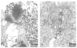
图1 (a)HIV-1 SH01大小不一的特征明显,核心颗粒呈锥形、杆状或圆形。(b)HIV-1 SH01从胞膜出芽释放过程的各个阶段均可见到,一即将脱落的HIV颗粒仍通过纤细的桥样连接与细胞膜相连。
Figure 1 (a) HIV-1 SH01 particles varied in size with conical, tubular or circular cores. (b) The whole budding process of HIV-1 SH01 was shown and an HIV particle was still connected to cell membrane by a fine bridge-like linkage.
, 百拇医药
本研究中,偶尔也可在HIV-1 SH01感染的MT4细胞的胞浆空泡内见到HIV颗粒(图2,a),并且在感染7天或持续感染的MT4细胞中均能见到。在有的空泡内,HIV颗粒还相当多见,且形态十分典型,不过除HIV颗粒外,还有细胞碎片和溶酶体结构(图2,b)。可见,此类空泡实际上是HIV的吞噬泡。在HIV-1持续感染的MT4转化细胞中,所见的一HIV吞噬泡位于细胞膜附近,可能是通过吞饮作用刚形成不久的吞饮泡(图2,c)。
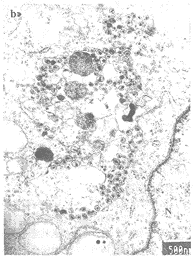
图2 (a)在HIV-1 SH01感染7天的MT4细胞中,一胞浆空泡内可见HIV颗粒和细胞碎片。(b)在同一标本的另一空泡中,存在大量的HIV颗粒,核心颗粒的锥形特征明显,同时溶酶体和细胞碎片亦清晰可见。(c)在HIV-1 SH01持续感染的MT4转化细胞中,一已摄入HIV颗粒的空泡位于细胞膜附近,可能是刚形成不久的吞饮泡。
, 百拇医药
Figure 2 (a) HIV particles and cell debris were found in a cytoplasmic vacuole in MT4 cells infected with HIV-1 SH01 at 7th day. (b) In the same HIV-1 SH01-infected MT4 cells, a lot of typical HIV particles were located in another cytoplasmic vacuole, lysosomes and cell debris could also be seen. (c) An HIV uptaking vacuole was situated at the edge of MT4 cells transformed persistently with HIV-1 SH01 and it is supposed that HIV particles might be engulfed by way of endocytosis.
, http://www.100md.com
另一罕见的现象是,溶酶体内大量HIV颗粒的存在。在HIV-1 SH01感染7天的MT4细胞中,偶然在一处观察到众多已摄入大量HIV颗粒的吞噬溶酶体,这些颗粒的形态特点与细胞外的HIV颗粒完全一致。这些吞噬了大量HIV颗粒的溶酶体,已进行过多次反复的融合(图3,a),而且部分初级溶酶体仍在继续与这些吞噬溶酶体进行融合(图3,b)。与上述的偶尔所见形成鲜明对比的是,在HIV-1 SH01持续感染的MT4转化细胞中,溶酶体摄取HIV颗粒的现象则相当普遍,尽管溶酶体内HIV颗粒的量要少一些(图4)。此外,还能见到HIV颗粒被消化、降解的过程,部分HIV颗粒的降解残体或碎片依稀可见。但同时在部分含HIV颗粒的吞噬溶酶体,中也存在溶酶体膜溶解的现象,不过其中的HIV颗粒已被基本降解或正被降解(图4)。结合前述的胞浆内HIV吞噬泡,细胞摄取HIV颗粒并经过溶酶体消化降解HIV的过程已不难理解。
图3 (a)在感染7天的HIV-1 SH01/MT4细胞中,众多的溶酶体已摄入大量的HIV颗粒,并已进行过多次反复的融合。(b)一些初级溶酶体仍在继续与已摄入大量HIV颗粒的吞噬溶酶体发生融合。
, 百拇医药
Figure 3 (a) In HIV-1 SH01-infected MT4 cells at 7th day, there were many phagolysosomes having taken in a lot of HIV particles and they had fused each other more than once. (b) Some primary lysosomes were continually fusing with these HIV uptaking phagolysosomes.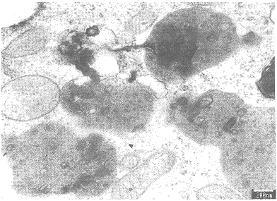
图4 在HIV-1 SH01持续感染的MT4转化细胞中,溶酶体摄入HIV颗粒的现象则相当普遍。在吞噬溶酶体内,部分HIV颗粒正被消化、降解,并可见其降解残体(“→”所示),但同时也存在着溶酶体膜溶解的现象(“▲”所示)。
Figure 4 HIV uptaking phagolysosomes were quite common in MT4 cells transformed persistently with HIV-1 SH01 and HIV particles seemed to be digested in some phagolysosomes and their remnants could also be seen (arrow) , but the dissolution of phagolysosome membrane existed there simultaneously (arrowhead).
, 百拇医药
此外,在HIV-1 SH01感染7天的MT4细胞的胞浆基质中,偶尔可观察到一种直径为80~200nm的病毒样颗粒。此颗粒大小不一的特征更为明显,呈椭圆形或圆形,其核心为圆形、椭圆形或长椭圆形,而非锥形,相对比例较大,轮廓欠清晰(图5,a)。在正常的MT4细胞中未能找到相同的颗粒(图5,b)。这些颗粒大多位于高尔基体附近,其形成可能与高尔基体有密切关系(图5,c)。有的尚未完成包被的病毒样颗粒与胞浆的某囊性结构仍然相连(图5,d);有的似乎正处于分泌过程中,而且其核心物质也同时进入此颗粒内(图5,e)。还直接可见一紧靠高尔基体分泌区的囊性结构分泌产生这种颗粒的过程(图5,f)。

图5 (a) 在HIV-1 SH01/MT4细胞(感染7天)的胞浆基质中可见一种病毒样颗粒,其大小不一的特征更为明显,核心颗粒为圆形或椭圆形,相对比例较大。(b)在正常的MT4细胞始终未能找到病毒样颗粒,其高尔基体区可见正常的分泌小泡。(c)此病毒样颗粒多位于高尔基体附近,其产生可能与高尔基体有关。(d)部分尚未完成包被的病毒样颗粒仍与胞浆的囊腔样结构相连接,可能由其延伸而来。(e)胞浆某囊性结构似正在分泌产生病毒样颗粒,其核心物质亦同时进入颗粒内(图2.c“→”所示)。(f)一病毒样颗粒正从紧靠高尔基体分泌区的一囊腔样结构出芽生成(图2 c“▲”所示)。
, 百拇医药
Figure 5 (a) Some virus-like particles were found in cytoplasmic matrix of HIV-1 SH01-infected MT4 cells at 7th day, which were more diversified in size with cores bigger, circular or oval but not conical. (b) No virus-like particle was demonstrated in normal MT4 cells besides some small bubbles in Golgi zone. (c) These particles were often located around Golgi apparatus where they were supposed to produce from. (d) An incompletely enveloped virus-like particle was still connected to a saccule-like structure. (e). Enlarged from (c) (arrow), two virus-like particles seemed to protrude from a saccule-like structure and their cores were packed into envelope at the same time. (f) Enlarged from (c) (arrowhead) , a virus-like particle was just budding from a saccule near the secreting area of Golgi apparatus.
, http://www.100md.com
讨论
HIV-1 SH01分离株对MT4、MT2等T细胞来源的细胞系具有良好的敏感性,并诱导产生融合多核巨细胞[14],故为合体细胞诱导型(SI)T细胞敏感株[15,16]。其形态学特征与以往报道的完全一致,具有典型的锥形核心,并且大小不一的特征相当明显[3-5]。胞膜芽生释放的过程十分典型,从新月形隆起到最后形成完整的病毒颗粒脱落的各个阶段均能得到很好的显示。刚释放到胞外的HIV颗粒,包膜常有刺样突起,核心颗粒常缺如,或呈环状或圆饼状,为未成熟的病毒颗粒,但在蛋白酶的作用下,不久即可完成其最后的成熟过程,并形成最具特征性的锥形核心颗粒[4,5,35]。
HIV除了最常见的以芽生的方式释放至细胞外的成熟过程外,根据宿主细胞的不同,亦可出芽至胞浆空泡内[2,6-13]。HIV-1单核一巨噬细胞敏感株,主要出芽至内质网腔或高尔基体囊腔成为胞浆空泡内病毒[6-9],一般认为这种空泡内的病毒可以逃避机体免疫系统的清除,因而巨噬细胞被认为是人体内HIV的庇护所和储存库[9,17,18]。不过,在一些非巨噬细胞系中有时也能见到类似的情形况[2,10-13]。我们所见的胞浆空泡内的HIV颗粒与上述情况不同,空泡内除了HIV颗粒外,还有细胞碎片和溶酶体,显然,此类HIV空泡很可能是通过吞饮作用从细胞外摄入HIV颗粒后形成的一种吞饮泡,而非扩张的高尔基体囊腔或内质网腔。
, http://www.100md.com
溶酶体摄入并消化HIV颗粒的现象曾有报道[2,9]。细胞外的HIV颗粒可通过与细胞膜融合或吞饮作用等方式进入细胞,后者形成为吞噬体,并与溶酶体发生融合[19-22]。我们所见的另一现象――HIV吞噬溶酶体,正是溶酶体与HIV吞噬体融合的结果。但许多研究表明,HIV与其他病毒(特别是流感病毒)一样,对溶酶体与吞噬体的融合具有抑制作用[9,23-26]。进一步研究证实,gp120同样可以抑制其融合作用,而可溶性CD4和抗CD4抗体则可阻止其抑制作用。可见溶酶体与吞噬体融合功能的异常是由于gp120与CD4相互作用的结果[26]。也有研究认为,被溶酶体摄入的HIV并不一定能被消化,因为在被降解之前已出现吞噬溶酶体膜的溶解[22,27]。我们在HIV-1 SH01持续感染的MT4转化细胞中发现,溶酶体在摄入并消化降解HIV颗粒的同时,也存在吞噬溶酶体膜溶解的现象,不过这些溶酶体中的HIV颗粒已被基本降解或正被降解。由此可见,溶酶体摄取并消化HIV的过程可能是相当复杂的。据报道,无症状的HIV感染者每天产生的病毒量平均达10.3×10⁹,远远超过人们原先的估计。只是人体的免疫系统对HIV同样具有强大的清除力,故而能维持相对的平衡而不发病,一旦这种较量向病毒倾斜,则发展为艾滋病即不可避免[28-30]。结合本研究的结果,我们认为,尽管特异性免疫对于维持HIV感染者的暂时不发病状态起主要作用,但非特异免疫在清除HIV方面的作用及重要性仍值得进一步研究和重视。
, http://www.100md.com
研究表明,某些HIV的gag基因突变株可改变原来胞膜芽生释放的途径为出芽至胞浆空泡内[31,32],不过这类变异株的核心颗粒均无锥形特征,也无感染力,是缺陷型的HIV病毒[33-36]。实际上,核心颗粒的形态是确定HIV是否成熟的最重要的标志[3-5];锥形核心既是HIV颗粒成熟的象征,也是其具有感染力的体现,因为只有具备这一特征的HIV毒株才能完成其复制的早期过程[35]。我们在胞浆基质中所观察到的病毒样颗粒,其大小与HIV颗粒相似,但核心颗粒的形态显然不同。因此我们推测,这种病毒样颗粒可能也是一种改变了释放途径的HIV缺陷病毒。不过,尽管在正常的MT4细胞中未能找到相同的颗粒,但从形态发生学看,由于这种颗粒很可能由高尔基体所分泌,因此尚不能排除前溶酶体的可能性,还需要作进一步的研究予以证实。
致谢:上海医科大学电镜教研室凌诒萍、吴正泉、俞永富教授,上海医科大学组胚教研室成令忠、陈红教授。
, http://www.100md.com
作者简介:李关汉(1962-),男,浙江遂昌人,副主任医师,硕士。曾从事小儿乙型肝炎和肝炎后肝硬化的临床、病理和免疫组织化学研究,后从事肠道病毒研究。1994年开始从事艾滋病毒的生物学特性和抗HIV中草药的筛选及有效成分的分离纯化等研究工作。现为中国预防医学科学院卫生部艾滋病预防控制中心临床病毒实验室在读博士生。
参考文献
1 Gelderblom H R, Ozel M, Pauli G. Morphogenesis and morphology of HIV structure-function relations [J] . Arch Virol, 1989, 106: 1-13.
2 Munn R J, Marx P A, Yamamoto J K, et al. Ultrastructural comparison of the retroviruses associated with human and simian acquired immunodeficiency syndromes [J] . Lab Invest, 1985, 53: 194-199.
, http://www.100md.com
3 Hockley D J. Wood R D, Jacobs J P, et al. Electron microscopy of human immunodeficiency virus [J] . J Gen Virol, 1988, 69: 2455-2649.
4 Katsumoto T, Hattori N, Kurimura T. Maturation of human imnunodeficiency virus, strain LAV, in vitro[J]. Intervirology, 1987, 27: 148-153.
5 Katsumoto T. Hattori N, Yamada O, et al. Intermediate virions in maturation of human immunodeficiency virus, strain LAV [J]. J Electron Microsc, 1988, 37: 205-207.
, http://www.100md.com
6 Gartner S, Markovits P, Markovitz D M, et al. The role of mononuclear phagocytes in HTLV-Ⅲ/LAV infection [J] . Science, 1986, 233: 215-218.
7 Geldelman H E, Orenstein J M, Martin M A, et al. Efficient isolation and propagation of human immunodeficiency virus on recombinant colony-stimulating factor l-treated monocytes [J]. J Exp Med, 1988, 167: 1428-1441.
8 Pautrat G, Suzan M, Aslaun D, et al. Human immunodeficiency virus type l infection of U937 cells promotes cell differentiation and a new pathway of viral assembly [J] . Virology, 1990, 179: 749-758.
, 百拇医药
9 Orenstein J M, Meltzer M S, Phipps T, et al. Cytoplasmic assembly and accumulation of human immunodeficiency virus types 1 and 2 in recombinant human colony-stimulating factor-l-treated human monocytes: an ultrastructural study [J] . J Virol, 1988, 62: 2578-2586.
10 Grief C, Farrar G H, Kent K A, et al. The assembly of HIV within the Golgi apparatus and Golgi-derived vesicles of JM cell syncytia [J]. AIDS, 1991, 5: 1433-1439.
, 百拇医药 11 Li Q, Zhang Y, Liang Y, et al. The morphogenesis of a Chinese strain of HIV-1 forming inclusion bodies in Jurkat-tatⅢ cells[J]. J Aquir Immune Defic Hum Retrovirol, 1995, 9: 103-113.
12 Klimkait T, Strebel K, Hoggan D, et al. The human immunodeficiency virus type 1-specific protein vpu is required for efficient virus maturation and release [J] . J Virol, 1990, 64: 621-629.
13 Srinivasan A, Goldsmith C S, York D, et al. Studies on human immunodeficiency virus-induced cytopathic effects [J] . Arch Virol, 1988, 99: 21-30.
, 百拇医药
14 李关汉,康来仪,金子辰,等.人类免疫缺陷病毒-1 SH01株的生物学特性研究[J].中华微生物学和免疫学杂志,1998,18:96-97.
15 Shioda T, Levy J A, Cheng-Mayer C. Macrophage and T cell-line tropisms of HIV-1 are determined by specific regions of the envelope gp120 gene [J] . Nature, 1991, 349: 167-169.
16 Schuitemaker H, Koot M, Kootstra N A, et al. Biologica1 phenotype of human immunodeficiency virus type 1 clones at different stages of infection: progression of disease is associated with a shift from monocytotropic to T-cell-tropic virus populations [J] . J Virol, 1992, 66: 1354-1360.
, http://www.100md.com
17 Schmitt M P, Steffan A M, Gendrault J L, et al. Multiplication of human immunodefiency virus in primary cultures of human kupffer cells-possible role of liver macrophage infection in the physiopathology of AIDS [J] . Res Virol, 1990, 141: 143-152.
18 Perelson A S, Essunger P, Cao Y, et al. Decay characteristics of HIV-1 infected components during combination therapy [J] . Nature, 1997, 387: 189-191.
19 Pauza C D, Price T M. Human immunodeficiency virus infection of T cells and monocytes proceeds via receptor-mediated endocytosis [J] . J Cell Biol, 1988, 107: 959-968.
, 百拇医药
20 Goto T, Harada S, Yamamoto N, et al. Entry of human immunodeficiency virus (HIV) into MT2, human T cell leukemia virus carrier cell line [J] . Arch Virol, 1988, 102: 29-38.
21 Nakai M and Goto T. Ultrastructure and morphogenesis of human immunodeficiency virus [J] . J Electron Microsc, 1996, 45: 247-257.
22 Phillips D M, Tan X. HIV-1 infection of the trophoblast cell line BeWo: a study of virus uptake [J] . AIDS Res Hum Retroviruses, 1992, 8: 1683-1691.
, 百拇医药
23 Baldwin G C, Fleischmann J, Chung Y, et al. Human immunodeficiency virus causes mononuclear phagocyte dysfunction [J]. Proc Natl Acad Sci USA, 1990, 87: 3933-3937.
24 Pittis M G, Sternik G, Sen L, et al. Impaired phagolysosomal fusion of peripheral blood monocytes from HIV-infected subjects [J]. Scand J Immunol, 1993, 38: 423-427.
25 Biggs B A, Hewish M, Kent S, et al. HIV-1 infection of human macrophages impairs phagocytosis and killing of toxoplasma gondii [J]. J Immunol, 1995, 154: 6132-6139.
, http://www.100md.com
26 Moorjani H, Craddock B P, Morrison S A, et al. Impairment of phagosome-lysosome fusion in HIV-1-infected macrophages [J] . J Aquir Immune Defic Hum Retrovirol, 1996, 13: 18-22.
27 Bourinbaiar A S, Phillips D M. Transmission of human immunodeficiency virus from monocytes to epithelia [J] . J Acquir Immune Defic Syndr, 1991, 4: 56-63.
28 Perelson A S, Neumann A U, Markowitz M, et al. HIV-1 dynamics in vivo: virion clearance rate, infected cell life-span, and viral generation time [J]. Science, 1996, 271: 1582-1585.
, 百拇医药
29 Levy J A. HIV pathogensis and long-term survival [J]. AIDS, 1993, 7: 1401-1410.
30 Pantaleo G, Fauci A S. Immunopathogenesis of HIV infection [J]. Annu Rev Microbiol, 1996, 50: 825-854.
31 Facke M, Janetzko A, Shoeman R L, et al. A large deletion in the matrix domain of the human immunodeficiency virus gag gene redirects virus particle assembly from the plasma membrane to the endoplasmic reticulum [J] . J Virol, 1993, 67: 4972-4980.
, 百拇医药
32 Freed E O, Orenstein J M, Bukler-White A J, et al. Single amino acid changes in the human immunodeficiency virus type 1 matrix protein block virus particle production [J] . J Virol, 1994, 68: 5311-5320.
33 Dorfman T, Bukovsky A, Ohagen A, et al. Functional domains of the capsid protein of human immunodeficiency virus type 1 [J] . J Virol, 1994, 68: 8180-8187.
34 Reicin A S, Berowitz P R, Luban J, et al. Linker insertion mutations in the human immunodeficiency virus type 1 gag gene: effects on virion particle assembly, release, and infectivity [J] . J Virol, 1995, 69: 642-650.
, http://www.100md.com
35 Reicin A S, Ohagen A, Yin L, et al. The role of gag in human immunodeficiency virus type 1 virion morphogenesis and early steps of the viral life cycle [J] , J Virol, 1996, 70: 8645-8652.
36 Ono A, Huang M, Freed E O. Characterization of human immunodeficiency virus type 1 matix revertants: effects on virus assembly, gag processing, and env incorporation into virions [J] . J Virol, 1997, 71: 4409-4418
收稿日期:1998-07-10;修回日期:1999-12-18, 百拇医药
单位:上海市疾病预防控制中心艾滋病毒实验室,上海市艾滋病监测中心,上海 200336
关键词:人免疫缺陷病毒1型(HIV-1)SH01株;形态发生学;吞噬溶酶体
病毒学报990403 摘要 用外周血单个核细胞混合培养法分离到的HIV-1 SH01株,具有典型的HIV颗粒的形态学特征,核心颗粒呈锥形,可见芽生释放的全过程。偶尔可在胞装空泡内见到HIV颗粒,同时还有细胞碎片和溶酶体结构,故此类空泡实际为HIV吞噬泡。另一少见的现象是溶酶体摄取并消化HIV颗粒,在HIV-1 SH01株感染7天或持续感染的MT4细胞中均可见到,后者尤为普遍。在HIV-1 SH01株持续感染的MT4转化细胞中,经过溶酶体消化的HIV残体依稀可见,但同时也存在吞噬溶酶体膜溶解的现象。可见,非特异性免疫在清除体内HIV方面的作用值得作进一步的研究。此外,在HIV-1 SH01株感染7天的MT4细胞的胞浆基质中,还可见到一种类病毒样颗粒,其性质有待进一步确定。
, 百拇医药
中图分类号:R512.91; Q934.2 文献标识码:A 文章编号:1000-8721(1999)04-0305-09
MORPHOLOGY AND MORPHOGENESIS OF HUMAN IMMUNODEFICIENCY VIRUS-1 ISOLATE, SH01 IN MT4 CELLS
LI Guan-han, WANG Mei-hua, JIN Zhi-cheng, PAN Qi-chao, KANG Lai-yi
(HIV Laboratory, Shanghai Center of Disease's Control and Prevention, Shanghai Municipal AIDS Surveillance Center, Shanghai 200336, China)
Abstract: An HIV-1 strain, SH01, isolated by coculture of peripheral blood mononuclear cells (PBMC) was easily passaged in MT4 cell line and multinuclear giant syncytia could be induced in MT2 cells. It was a syncytium-inducing (SI) T-cell-tropic isolate. Electron microscopy showed this isolate shared the typical features of HIV-1 in morphology, such as conical core, variation in size and budding release from cell membrane. A distinct fact was that HIV particles could be found in cytoplasmic vacuoles in MT4 cells infected at 7th day or trasformed persistently with HIV-1 SH01, but cell debris or lysosomes were seen at the same time. So these vacuoles were phagosomes or endocytic vacuoles rather than dilated vesicles of Golgi apparatus or endoplasmic reticulum. Furthermore, a large number of HIV particles were also found by chance in phagolysosomes in HIV-1 SH01-infected MT4 cells at 7th day, but it was quite common in HIV-1 SH01-transformed MT4 cells and the remnants of HIV particles were located in some phagolysosomes, yet the dissolution of phagolysosome membrane could be demonstrated simultaneously. Conclusively, it is evident that HIV particles were taken in by MT4 cells through endocytosis and digested by lysosomes, so the role of lysosomes should be paid attention to more extensively in the clearance of HIV in human body. In addition, a kind of virus-like particles was located in cytoplasmic matrix rather than in vacuoles or vesicles, which were more diversified in size with cores bigger, circular or oval and possibly produced from Golgi apparatus, but their characteristics needs to be further determined.
, 百拇医药
Key words: Human immunodeficiency virus-1 (HIV-1) SH01; Morphogenesis; Phagolysosome
人类免疫缺陷病毒(HIV)是慢病毒属(lentivirus)中典型的C型病毒,与同属的其他病毒比较,其非对称的锥形核心颗粒为其最重要的形态学特征之一[1,2]。形态发生学研究表明,HIV主要以芽生的方式释放至细胞外,刚从细胞膜脱落的HIV颗粒尚无典型的锥形核心颗粒,核心颗粒的成熟是在释放之后较短的时间内完成的[1-5]。但是,根据宿主细胞的不同,HIV亦可释放至胞浆的内质网腔或高尔基体腔,成为空泡内病毒[2,6-13],甚至于形成包涵体[11]。本实验室于1994年底至1995年初从本市的一名曾去非洲莱索托从事劳务活动的HIV-1感染者中分离到一株病毒,即HIV-1 SH01,为合体细胞诱导型(SI)T细胞敏感株[14-16]。超微结构和形态发生学研究显示,此毒株在MT4细胞除具有HIV-1的共同特征外,还可观察到HIV的吞噬泡及溶酶体摄取并消化HIV颗粒的现象,在胞浆基质中尚可观察到一种病毒样颗粒。
, http://www.100md.com
材料与方法
1 HIV-1的分离及检测
HIV-1的分离:从一名曾在莱索托从事劳务活动一年半的HIV-1感染者中抽取新鲜血液10ml,加肝素(20单位/ml)抗凝。经Hank’s液双倍稀释后,以Ficoll-Hypaque液分离并收集外周血单个核细胞(PBMC),然后与经PHA刺激3天的正常PBMC混合。在含20%NCS、10%~20%IL-2和抗菌素的RPMI 1640中继续培养。每周补充PHA激活的正常PBMC1次,换液2次。 间接免疫荧光法(IFA):常规制备细胞涂片,以HIV-1阳性血清为一抗,FITC兔抗人IgG(Dako公司)为二抗,进行常规荧光染色并观察。
HIV-1 P24抗原测定:采用Vironstika半定量ELISA试剂盒测定PBMC共培养上清的HIV-1 P24抗原。
蛋白印迹法(Western blot):HIV-1毒株的裂解抗原,经聚丙烯酰胺梯度凝胶电泳后,将抗原转移至硝酸纤维膜,并以5%脱脂奶作封闭。转移膜相继在1:100 HIV-1阳性血清及1:100 HRP羊抗人IgG中孵育,DAB-H2O2显色,清水中止反应。
, http://www.100md.com
2 HIV-1分离株的转移和传代 将PBMC共培养上清反复感染MT4、H9、Molt4/clone8细胞,并做IFA。HIV-1分离株转移到MT4细胞之后,继续换液培养,使少量存活细胞成为HIV-1持续感染的转化细胞。将该毒株感染的MT4细胞培养液再感染H9、Molt4/clone8和MT2等细胞,并观察各种细胞的病变情况。
3 电境观察 取HIV-1分离株感染7天的MT4细胞、持续感染的MT4转化细胞和正常MT4细胞悬液各10ml,置尖底管中,以4 000r/min离心10分钟,去上清。在4℃经2.5%戊二醛固定24小时后,以1%锇酸后固定。乙醇-丙酮梯度脱水,618环氧树脂包埋,制超薄切片,以醋酸双氧铀和枸橼酸铅染色,在JEM-1200EX透射电镜中观察。
结果
建立PBMC共培养之后,每周进行IFA染色,并检测共培养上清的P24抗原。第22天P24抗原阳性,第6周IFA染色明显阳性,此分离株即HIV-1 SH01。将共培养上清反复感染MT4等细胞,5周后此毒株成功转移至MT4细胞,并出现细胞病变,表现为细胞固缩、崩解和气球样细胞。继续培养HIV-1 SH01/MT4细胞,少数未完全病变的细胞逐渐恢复正常形态,成为此毒株持续感染的转化细胞。将HIV-1 SH01/MT4细胞的培养上清感染H9、Molt4/clone8、MT2和CEM-ss细胞,除H9细胞外,其他细胞均相当敏感。蛋白印迹法分析显示,该毒株的抗原特征与HIV-1 Ⅲb基本一致[14]。
, http://www.100md.com
HIV-1 SH01具有HIV的典型形态特征,呈圆形或椭圆形,核心颗粒为锥形,根据切面的不同亦可表现为杆状或圆形。病毒颗粒直径一般在80~150nm之间,最大的达175nm,大小不一的特征明显(图1,a)。可观察到典型的HIV颗粒自胞膜芽生释放的全过程。出芽处胞膜起初呈新月形隆起,逐渐成半圆形、圆形,最后成完整的病毒颗粒而脱落,有的即将脱落的病毒颗粒仍通过纤细的桥样连接与细胞膜相连(图1,b)。刚释放到细胞外的病毒颗粒外膜有刺样突起,核心颗粒常缺如,此为未成熟的HIV颗粒。
图1 (a)HIV-1 SH01大小不一的特征明显,核心颗粒呈锥形、杆状或圆形。(b)HIV-1 SH01从胞膜出芽释放过程的各个阶段均可见到,一即将脱落的HIV颗粒仍通过纤细的桥样连接与细胞膜相连。
Figure 1 (a) HIV-1 SH01 particles varied in size with conical, tubular or circular cores. (b) The whole budding process of HIV-1 SH01 was shown and an HIV particle was still connected to cell membrane by a fine bridge-like linkage.
, 百拇医药
本研究中,偶尔也可在HIV-1 SH01感染的MT4细胞的胞浆空泡内见到HIV颗粒(图2,a),并且在感染7天或持续感染的MT4细胞中均能见到。在有的空泡内,HIV颗粒还相当多见,且形态十分典型,不过除HIV颗粒外,还有细胞碎片和溶酶体结构(图2,b)。可见,此类空泡实际上是HIV的吞噬泡。在HIV-1持续感染的MT4转化细胞中,所见的一HIV吞噬泡位于细胞膜附近,可能是通过吞饮作用刚形成不久的吞饮泡(图2,c)。
图2 (a)在HIV-1 SH01感染7天的MT4细胞中,一胞浆空泡内可见HIV颗粒和细胞碎片。(b)在同一标本的另一空泡中,存在大量的HIV颗粒,核心颗粒的锥形特征明显,同时溶酶体和细胞碎片亦清晰可见。(c)在HIV-1 SH01持续感染的MT4转化细胞中,一已摄入HIV颗粒的空泡位于细胞膜附近,可能是刚形成不久的吞饮泡。
, 百拇医药
Figure 2 (a) HIV particles and cell debris were found in a cytoplasmic vacuole in MT4 cells infected with HIV-1 SH01 at 7th day. (b) In the same HIV-1 SH01-infected MT4 cells, a lot of typical HIV particles were located in another cytoplasmic vacuole, lysosomes and cell debris could also be seen. (c) An HIV uptaking vacuole was situated at the edge of MT4 cells transformed persistently with HIV-1 SH01 and it is supposed that HIV particles might be engulfed by way of endocytosis.
, http://www.100md.com
另一罕见的现象是,溶酶体内大量HIV颗粒的存在。在HIV-1 SH01感染7天的MT4细胞中,偶然在一处观察到众多已摄入大量HIV颗粒的吞噬溶酶体,这些颗粒的形态特点与细胞外的HIV颗粒完全一致。这些吞噬了大量HIV颗粒的溶酶体,已进行过多次反复的融合(图3,a),而且部分初级溶酶体仍在继续与这些吞噬溶酶体进行融合(图3,b)。与上述的偶尔所见形成鲜明对比的是,在HIV-1 SH01持续感染的MT4转化细胞中,溶酶体摄取HIV颗粒的现象则相当普遍,尽管溶酶体内HIV颗粒的量要少一些(图4)。此外,还能见到HIV颗粒被消化、降解的过程,部分HIV颗粒的降解残体或碎片依稀可见。但同时在部分含HIV颗粒的吞噬溶酶体,中也存在溶酶体膜溶解的现象,不过其中的HIV颗粒已被基本降解或正被降解(图4)。结合前述的胞浆内HIV吞噬泡,细胞摄取HIV颗粒并经过溶酶体消化降解HIV的过程已不难理解。
图3 (a)在感染7天的HIV-1 SH01/MT4细胞中,众多的溶酶体已摄入大量的HIV颗粒,并已进行过多次反复的融合。(b)一些初级溶酶体仍在继续与已摄入大量HIV颗粒的吞噬溶酶体发生融合。
, 百拇医药
Figure 3 (a) In HIV-1 SH01-infected MT4 cells at 7th day, there were many phagolysosomes having taken in a lot of HIV particles and they had fused each other more than once. (b) Some primary lysosomes were continually fusing with these HIV uptaking phagolysosomes.
图4 在HIV-1 SH01持续感染的MT4转化细胞中,溶酶体摄入HIV颗粒的现象则相当普遍。在吞噬溶酶体内,部分HIV颗粒正被消化、降解,并可见其降解残体(“→”所示),但同时也存在着溶酶体膜溶解的现象(“▲”所示)。
Figure 4 HIV uptaking phagolysosomes were quite common in MT4 cells transformed persistently with HIV-1 SH01 and HIV particles seemed to be digested in some phagolysosomes and their remnants could also be seen (arrow) , but the dissolution of phagolysosome membrane existed there simultaneously (arrowhead).
, 百拇医药
此外,在HIV-1 SH01感染7天的MT4细胞的胞浆基质中,偶尔可观察到一种直径为80~200nm的病毒样颗粒。此颗粒大小不一的特征更为明显,呈椭圆形或圆形,其核心为圆形、椭圆形或长椭圆形,而非锥形,相对比例较大,轮廓欠清晰(图5,a)。在正常的MT4细胞中未能找到相同的颗粒(图5,b)。这些颗粒大多位于高尔基体附近,其形成可能与高尔基体有密切关系(图5,c)。有的尚未完成包被的病毒样颗粒与胞浆的某囊性结构仍然相连(图5,d);有的似乎正处于分泌过程中,而且其核心物质也同时进入此颗粒内(图5,e)。还直接可见一紧靠高尔基体分泌区的囊性结构分泌产生这种颗粒的过程(图5,f)。
图5 (a) 在HIV-1 SH01/MT4细胞(感染7天)的胞浆基质中可见一种病毒样颗粒,其大小不一的特征更为明显,核心颗粒为圆形或椭圆形,相对比例较大。(b)在正常的MT4细胞始终未能找到病毒样颗粒,其高尔基体区可见正常的分泌小泡。(c)此病毒样颗粒多位于高尔基体附近,其产生可能与高尔基体有关。(d)部分尚未完成包被的病毒样颗粒仍与胞浆的囊腔样结构相连接,可能由其延伸而来。(e)胞浆某囊性结构似正在分泌产生病毒样颗粒,其核心物质亦同时进入颗粒内(图2.c“→”所示)。(f)一病毒样颗粒正从紧靠高尔基体分泌区的一囊腔样结构出芽生成(图2 c“▲”所示)。
, 百拇医药
Figure 5 (a) Some virus-like particles were found in cytoplasmic matrix of HIV-1 SH01-infected MT4 cells at 7th day, which were more diversified in size with cores bigger, circular or oval but not conical. (b) No virus-like particle was demonstrated in normal MT4 cells besides some small bubbles in Golgi zone. (c) These particles were often located around Golgi apparatus where they were supposed to produce from. (d) An incompletely enveloped virus-like particle was still connected to a saccule-like structure. (e). Enlarged from (c) (arrow), two virus-like particles seemed to protrude from a saccule-like structure and their cores were packed into envelope at the same time. (f) Enlarged from (c) (arrowhead) , a virus-like particle was just budding from a saccule near the secreting area of Golgi apparatus.
, http://www.100md.com
讨论
HIV-1 SH01分离株对MT4、MT2等T细胞来源的细胞系具有良好的敏感性,并诱导产生融合多核巨细胞[14],故为合体细胞诱导型(SI)T细胞敏感株[15,16]。其形态学特征与以往报道的完全一致,具有典型的锥形核心,并且大小不一的特征相当明显[3-5]。胞膜芽生释放的过程十分典型,从新月形隆起到最后形成完整的病毒颗粒脱落的各个阶段均能得到很好的显示。刚释放到胞外的HIV颗粒,包膜常有刺样突起,核心颗粒常缺如,或呈环状或圆饼状,为未成熟的病毒颗粒,但在蛋白酶的作用下,不久即可完成其最后的成熟过程,并形成最具特征性的锥形核心颗粒[4,5,35]。
HIV除了最常见的以芽生的方式释放至细胞外的成熟过程外,根据宿主细胞的不同,亦可出芽至胞浆空泡内[2,6-13]。HIV-1单核一巨噬细胞敏感株,主要出芽至内质网腔或高尔基体囊腔成为胞浆空泡内病毒[6-9],一般认为这种空泡内的病毒可以逃避机体免疫系统的清除,因而巨噬细胞被认为是人体内HIV的庇护所和储存库[9,17,18]。不过,在一些非巨噬细胞系中有时也能见到类似的情形况[2,10-13]。我们所见的胞浆空泡内的HIV颗粒与上述情况不同,空泡内除了HIV颗粒外,还有细胞碎片和溶酶体,显然,此类HIV空泡很可能是通过吞饮作用从细胞外摄入HIV颗粒后形成的一种吞饮泡,而非扩张的高尔基体囊腔或内质网腔。
, http://www.100md.com
溶酶体摄入并消化HIV颗粒的现象曾有报道[2,9]。细胞外的HIV颗粒可通过与细胞膜融合或吞饮作用等方式进入细胞,后者形成为吞噬体,并与溶酶体发生融合[19-22]。我们所见的另一现象――HIV吞噬溶酶体,正是溶酶体与HIV吞噬体融合的结果。但许多研究表明,HIV与其他病毒(特别是流感病毒)一样,对溶酶体与吞噬体的融合具有抑制作用[9,23-26]。进一步研究证实,gp120同样可以抑制其融合作用,而可溶性CD4和抗CD4抗体则可阻止其抑制作用。可见溶酶体与吞噬体融合功能的异常是由于gp120与CD4相互作用的结果[26]。也有研究认为,被溶酶体摄入的HIV并不一定能被消化,因为在被降解之前已出现吞噬溶酶体膜的溶解[22,27]。我们在HIV-1 SH01持续感染的MT4转化细胞中发现,溶酶体在摄入并消化降解HIV颗粒的同时,也存在吞噬溶酶体膜溶解的现象,不过这些溶酶体中的HIV颗粒已被基本降解或正被降解。由此可见,溶酶体摄取并消化HIV的过程可能是相当复杂的。据报道,无症状的HIV感染者每天产生的病毒量平均达10.3×10⁹,远远超过人们原先的估计。只是人体的免疫系统对HIV同样具有强大的清除力,故而能维持相对的平衡而不发病,一旦这种较量向病毒倾斜,则发展为艾滋病即不可避免[28-30]。结合本研究的结果,我们认为,尽管特异性免疫对于维持HIV感染者的暂时不发病状态起主要作用,但非特异免疫在清除HIV方面的作用及重要性仍值得进一步研究和重视。
, http://www.100md.com
研究表明,某些HIV的gag基因突变株可改变原来胞膜芽生释放的途径为出芽至胞浆空泡内[31,32],不过这类变异株的核心颗粒均无锥形特征,也无感染力,是缺陷型的HIV病毒[33-36]。实际上,核心颗粒的形态是确定HIV是否成熟的最重要的标志[3-5];锥形核心既是HIV颗粒成熟的象征,也是其具有感染力的体现,因为只有具备这一特征的HIV毒株才能完成其复制的早期过程[35]。我们在胞浆基质中所观察到的病毒样颗粒,其大小与HIV颗粒相似,但核心颗粒的形态显然不同。因此我们推测,这种病毒样颗粒可能也是一种改变了释放途径的HIV缺陷病毒。不过,尽管在正常的MT4细胞中未能找到相同的颗粒,但从形态发生学看,由于这种颗粒很可能由高尔基体所分泌,因此尚不能排除前溶酶体的可能性,还需要作进一步的研究予以证实。
致谢:上海医科大学电镜教研室凌诒萍、吴正泉、俞永富教授,上海医科大学组胚教研室成令忠、陈红教授。
, http://www.100md.com
作者简介:李关汉(1962-),男,浙江遂昌人,副主任医师,硕士。曾从事小儿乙型肝炎和肝炎后肝硬化的临床、病理和免疫组织化学研究,后从事肠道病毒研究。1994年开始从事艾滋病毒的生物学特性和抗HIV中草药的筛选及有效成分的分离纯化等研究工作。现为中国预防医学科学院卫生部艾滋病预防控制中心临床病毒实验室在读博士生。
参考文献
1 Gelderblom H R, Ozel M, Pauli G. Morphogenesis and morphology of HIV structure-function relations [J] . Arch Virol, 1989, 106: 1-13.
2 Munn R J, Marx P A, Yamamoto J K, et al. Ultrastructural comparison of the retroviruses associated with human and simian acquired immunodeficiency syndromes [J] . Lab Invest, 1985, 53: 194-199.
, http://www.100md.com
3 Hockley D J. Wood R D, Jacobs J P, et al. Electron microscopy of human immunodeficiency virus [J] . J Gen Virol, 1988, 69: 2455-2649.
4 Katsumoto T, Hattori N, Kurimura T. Maturation of human imnunodeficiency virus, strain LAV, in vitro[J]. Intervirology, 1987, 27: 148-153.
5 Katsumoto T. Hattori N, Yamada O, et al. Intermediate virions in maturation of human immunodeficiency virus, strain LAV [J]. J Electron Microsc, 1988, 37: 205-207.
, http://www.100md.com
6 Gartner S, Markovits P, Markovitz D M, et al. The role of mononuclear phagocytes in HTLV-Ⅲ/LAV infection [J] . Science, 1986, 233: 215-218.
7 Geldelman H E, Orenstein J M, Martin M A, et al. Efficient isolation and propagation of human immunodeficiency virus on recombinant colony-stimulating factor l-treated monocytes [J]. J Exp Med, 1988, 167: 1428-1441.
8 Pautrat G, Suzan M, Aslaun D, et al. Human immunodeficiency virus type l infection of U937 cells promotes cell differentiation and a new pathway of viral assembly [J] . Virology, 1990, 179: 749-758.
, 百拇医药
9 Orenstein J M, Meltzer M S, Phipps T, et al. Cytoplasmic assembly and accumulation of human immunodeficiency virus types 1 and 2 in recombinant human colony-stimulating factor-l-treated human monocytes: an ultrastructural study [J] . J Virol, 1988, 62: 2578-2586.
10 Grief C, Farrar G H, Kent K A, et al. The assembly of HIV within the Golgi apparatus and Golgi-derived vesicles of JM cell syncytia [J]. AIDS, 1991, 5: 1433-1439.
, 百拇医药 11 Li Q, Zhang Y, Liang Y, et al. The morphogenesis of a Chinese strain of HIV-1 forming inclusion bodies in Jurkat-tatⅢ cells[J]. J Aquir Immune Defic Hum Retrovirol, 1995, 9: 103-113.
12 Klimkait T, Strebel K, Hoggan D, et al. The human immunodeficiency virus type 1-specific protein vpu is required for efficient virus maturation and release [J] . J Virol, 1990, 64: 621-629.
13 Srinivasan A, Goldsmith C S, York D, et al. Studies on human immunodeficiency virus-induced cytopathic effects [J] . Arch Virol, 1988, 99: 21-30.
, 百拇医药
14 李关汉,康来仪,金子辰,等.人类免疫缺陷病毒-1 SH01株的生物学特性研究[J].中华微生物学和免疫学杂志,1998,18:96-97.
15 Shioda T, Levy J A, Cheng-Mayer C. Macrophage and T cell-line tropisms of HIV-1 are determined by specific regions of the envelope gp120 gene [J] . Nature, 1991, 349: 167-169.
16 Schuitemaker H, Koot M, Kootstra N A, et al. Biologica1 phenotype of human immunodeficiency virus type 1 clones at different stages of infection: progression of disease is associated with a shift from monocytotropic to T-cell-tropic virus populations [J] . J Virol, 1992, 66: 1354-1360.
, http://www.100md.com
17 Schmitt M P, Steffan A M, Gendrault J L, et al. Multiplication of human immunodefiency virus in primary cultures of human kupffer cells-possible role of liver macrophage infection in the physiopathology of AIDS [J] . Res Virol, 1990, 141: 143-152.
18 Perelson A S, Essunger P, Cao Y, et al. Decay characteristics of HIV-1 infected components during combination therapy [J] . Nature, 1997, 387: 189-191.
19 Pauza C D, Price T M. Human immunodeficiency virus infection of T cells and monocytes proceeds via receptor-mediated endocytosis [J] . J Cell Biol, 1988, 107: 959-968.
, 百拇医药
20 Goto T, Harada S, Yamamoto N, et al. Entry of human immunodeficiency virus (HIV) into MT2, human T cell leukemia virus carrier cell line [J] . Arch Virol, 1988, 102: 29-38.
21 Nakai M and Goto T. Ultrastructure and morphogenesis of human immunodeficiency virus [J] . J Electron Microsc, 1996, 45: 247-257.
22 Phillips D M, Tan X. HIV-1 infection of the trophoblast cell line BeWo: a study of virus uptake [J] . AIDS Res Hum Retroviruses, 1992, 8: 1683-1691.
, 百拇医药
23 Baldwin G C, Fleischmann J, Chung Y, et al. Human immunodeficiency virus causes mononuclear phagocyte dysfunction [J]. Proc Natl Acad Sci USA, 1990, 87: 3933-3937.
24 Pittis M G, Sternik G, Sen L, et al. Impaired phagolysosomal fusion of peripheral blood monocytes from HIV-infected subjects [J]. Scand J Immunol, 1993, 38: 423-427.
25 Biggs B A, Hewish M, Kent S, et al. HIV-1 infection of human macrophages impairs phagocytosis and killing of toxoplasma gondii [J]. J Immunol, 1995, 154: 6132-6139.
, http://www.100md.com
26 Moorjani H, Craddock B P, Morrison S A, et al. Impairment of phagosome-lysosome fusion in HIV-1-infected macrophages [J] . J Aquir Immune Defic Hum Retrovirol, 1996, 13: 18-22.
27 Bourinbaiar A S, Phillips D M. Transmission of human immunodeficiency virus from monocytes to epithelia [J] . J Acquir Immune Defic Syndr, 1991, 4: 56-63.
28 Perelson A S, Neumann A U, Markowitz M, et al. HIV-1 dynamics in vivo: virion clearance rate, infected cell life-span, and viral generation time [J]. Science, 1996, 271: 1582-1585.
, 百拇医药
29 Levy J A. HIV pathogensis and long-term survival [J]. AIDS, 1993, 7: 1401-1410.
30 Pantaleo G, Fauci A S. Immunopathogenesis of HIV infection [J]. Annu Rev Microbiol, 1996, 50: 825-854.
31 Facke M, Janetzko A, Shoeman R L, et al. A large deletion in the matrix domain of the human immunodeficiency virus gag gene redirects virus particle assembly from the plasma membrane to the endoplasmic reticulum [J] . J Virol, 1993, 67: 4972-4980.
, 百拇医药
32 Freed E O, Orenstein J M, Bukler-White A J, et al. Single amino acid changes in the human immunodeficiency virus type 1 matrix protein block virus particle production [J] . J Virol, 1994, 68: 5311-5320.
33 Dorfman T, Bukovsky A, Ohagen A, et al. Functional domains of the capsid protein of human immunodeficiency virus type 1 [J] . J Virol, 1994, 68: 8180-8187.
34 Reicin A S, Berowitz P R, Luban J, et al. Linker insertion mutations in the human immunodeficiency virus type 1 gag gene: effects on virion particle assembly, release, and infectivity [J] . J Virol, 1995, 69: 642-650.
, http://www.100md.com
35 Reicin A S, Ohagen A, Yin L, et al. The role of gag in human immunodeficiency virus type 1 virion morphogenesis and early steps of the viral life cycle [J] , J Virol, 1996, 70: 8645-8652.
36 Ono A, Huang M, Freed E O. Characterization of human immunodeficiency virus type 1 matix revertants: effects on virus assembly, gag processing, and env incorporation into virions [J] . J Virol, 1997, 71: 4409-4418
收稿日期:1998-07-10;修回日期:1999-12-18, 百拇医药