人升主动脉-主动脉弓的几何形态与显微结构
作者:蔡国君 姜宗来 纪荣明
单位:蔡国君 姜宗来 纪荣明(第二军医大学解剖学教研室,生物医学工程研究所,上海 200433)
关键词:主动脉弓;几何形态;血管平滑肌;弹性纤维;胶原纤维;生物力学
解剖学报000120 摘 要:目的 为主动脉弓生物力学和血流动力学研究提供形态学基础。 方法 应用组织学和计算机图像分析方法,对5例正常成人升主动脉-主动脉弓进行计量形态学研究。 结果 获得了人升主动脉-主动脉弓几何形态和显微结构成分含量沿轴向和周向连续变化的完整数据。升主动脉根部结构成分以胶原纤维为主,左、右、后瓣环、窦壁的厚度和结构成分含量在血管周向无显著差异。 结论 人主动脉弓几何形态与显微结构存在血管轴向和周向非均匀性,提示主动脉弓应力应变的不均匀状况。主动脉弓胶原纤维的周向非均匀性不如弹性纤维和平滑肌显著,与其力学性质相符合。
, 百拇医药
分类号:R322.1+21 文献标识码:A
文章编号:0529-1356(2000)01-82
GEOMETRICAL MORPHOLOGY AND MICROSTRUCTURE OF THE
HUMAN AORTIC ARCH
CAI Guo-jun
(Department of Anatomy,Institute of Biomedical Engineering,The Second Military Medical University,Shanghai 200433,China)
JIANG Zong-lai
, 百拇医药
(Department of Anatomy,Institute of Biomedical Engineering,The Second Military Medical University,Shanghai 200433,China)
JI Rong-ming
(Department of Anatomy,Institute of Biomedical Engineering,The Second Military Medical University,Shanghai 200433,China)
Abstract:Objective The aim of the present study is to provide essential morphological data for research concerning biomechanics and hemodynamics of the vessel.Methods A quantitative study was conducted on 5 ascending aorta and aortic arch specimens of normal adult human cadaver by continuous vascular sections and computer image analysis.Results A series of data were gained of geometrical morphology and relative area(Aa%) of smooth muscle cells,elastin and collagen in axial and circumferential directions of the ascending aorta and aortic arch.At the site of aortic root collagen is the main component.The difference of wall thickness and Aa% of the microstructural components among aortic rings and walls of aortic sinuses are all not significant.Conclusion The geometrical morphology and microstructure alter unequally in axial and circumferential directions of the aortic arch,which reflects the inequality of the vascular stress and strain.The inequality of collagen in circumferential directions is not as significant as that of elastin and smooth muscle cells,according with whose mechanical properties of high elastic modulus.
, http://www.100md.com
Key words:Aortic arch; Geometrical morphology; Smooth muscle cells; Elastin; Collagen; Biomechanics▲
升主动脉-主动脉弓是一段弯曲、分支的血管,其形态结构和血流动力学复杂。Wasano[1]和Sans[2]等对其做过不同侧重的研究,但有关升主动脉-主动脉弓几何形态和显微结构的定量资料尚不完整,对其结构的血管周向和轴向非均匀性未见报道。我们对正常成人升主动脉-主动脉弓进行了系统定量的形态学研究,为其生物力学和血流动力学研究提供必要的形态学基础,为主动脉瘤、动脉粥样硬化等主动脉常见疾病的临床诊治提供形态学依据。
材料和方法
1.取材方法和组织切片
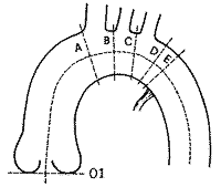
主动脉(包括升主动脉、主动脉弓和部分胸主动脉)新鲜标本5例,均为青年男性,生前无心血管相关疾病史。120mmHg压力下灌注固定后,自主动脉瓣环基底平面起,沿血管轴线量取升主动脉-主动脉弓全长150mm。垂直血管长轴,于A、B、C、D、E各平面处切断血管取材(图1),其余自主动脉瓣环基底平面至平面A血管段和平面E远段血管,分别按血管轴线长度平均7等分和4等分切断取材。标记血管段方位,以血管弓凸缘为上,凹缘为下,与上、下相垂直的方向,近腹侧为前,近背侧为后。常规石蜡切片,厚4μm。自主动脉瓣环基底平面起,各段切面分别标记为01、02……16。根据Han[3]的方法,将升主动脉-主动脉弓全长记为L,各切面距瓣环基底平面沿血管轴线的长度记为x,这样,主动脉上任一切面的位置可以用x/L表示。
, http://www.100md.com
图1 人升主动脉-主动脉弓模式图。示取材部位:
01:主动脉瓣环基底平面
A:头臂动脉起始处近端(08)
B:左颈总动脉起始处近端(09)
C:左锁骨下动脉起始处近端(10)
D:主动脉狭部中点(11)
E:动脉韧带附着处近端(12)
Fig.1 Diagram of the human ascending aorta and aortic arch,showing the special sections where measurements were taken:
, 百拇医药
01,Base attachment of the aortic root.
A,Proximal to the origin of the innominate artery(08).
B,Proximal to the origin of the left common carotid
artery (09).
C,Proximal to the origin of the left subclavian artery(10).
D,Middle point of the aortic isthmus(11).
E,Proximal to the anastomosis of the arterial ligament with
, http://www.100md.com
the aorta(12).
2.切片染色和图像分析
各血管段连续3张切片分别用Neubert酸性茜素蓝法、Weigert间苯二酚复红法和苯胺蓝法染血管平滑肌、弹性纤维和胶原纤维。光镜下用计算机图像分析测定血管的形态学指标。结果经统计学处理,两组资料比较用t检验,多组资料比较用方差分析。
结 果
1.升主动脉根部的几何形态与显微结构
在主动脉瓣环基底平面(切面01),胶原纤维与弹性纤维含量的比值(C/E值)显著大于主动脉其他部位(图2),血管平滑肌、弹性纤维和胶原纤维的相对含量在3个瓣环间均无显著差异,左、右、后3个瓣环的壁厚分别为497.0±22.7μm、508.1±26.3μm和499.9±25.7μm,三者无显著差异(图3)。
, http://www.100md.com
图2 人升主动脉-主动脉弓的C/E值。主动脉根部(切面01,02)的C/E值显著大于主动脉其他部位(P<0.01)
Fig.2 The C/E of the human ascending aorta and aortic arch.The C/E of the aortic root(section 01,02) is significantly larger than that of the other part of the aorta (P<0.01)
图3 主动脉瓣环基底平面(切面01)切片。L:左瓣环,R:右瓣环,P:后瓣环。
Fig.3 Cross-sectional view of the base attachment of the aortic root(section 01).L,left aortic ring;R,right aortic ring; P,posterior aortic ring.The difference of wall thickness among three aortic rings is not significant.
, http://www.100md.com
在主动脉窦平面(切面02),左、右、后窦壁厚度分别为624.6±29.7μm、631.2±33.4μm和627.5±30.6μm,三者无显著差异。窦壁为瓣环向主动脉壁的延续部分,其显微结构成分含量也界于两者之间,仍以胶原纤维为主,各显微结构成分的相对含量在3个窦壁间也无显著差异。
2.升主动脉-主动脉弓的几何形态学
升主动脉-主动脉弓的管径、壁厚、管壁和管腔横截面积在血管轴向连续变化(图4)。

轴向位置(x-L)
Axial location
图4 升主动脉-主动脉弓的几何形态在血管轴向的变化。
, 百拇医药
Fig.4 Line graphs show the geometrical morphology alter continuously and unequally along the
axis of the ascending aorta and aortic arch.
表1列出自升主动脉管部(切面03)起各血管切面在不同周向的中膜厚。其中在切面04~06、08~12和切面14等部位,中膜厚在周向呈显著非均匀性(图5,6),其在不同周向的两两比较见表2。
图5 升主动脉中段切片(切面04)。示血管中膜厚周向非均匀性,S:上壁,A:前壁,I:下壁,P:后壁。
Fig.5 Cross-sectional view of the middle part of the ascending aorta(section 04),showing the media thickness is unequal in different circumferential directions.S,superior; A,anterior; I,inferior,P,posterior.
, http://www.100md.com
图6 主动脉弓切面A切片。示血管中膜厚周向非均匀性,S:上壁,A:前壁,I:下壁,P:后壁。
Fig.6 Cross-sectional view of the aortic arch(section A),showing the media thickness is unequal in different circumferential directions.S,superior;A,anterior;I,inferior;P,posterior.
表1 升主动脉-主动脉弓各切面不同周向方向的中膜厚(μm, ±s)
±s)
Table 1 Media thickness of aorta in differentcircumferential directions(μm, ±s) 切面
, 百拇医药
section
上
superior
前
anterior
下
inferior
后
posterior
03
768.4±56.3
727.6±52.1
733.6±53.3
, 百拇医药
706.1±44.3
04
831.5±70.8
734.1±51.6
799.9±61.0
747.6±64.7**
05
833.4±51.6
741.3±46.1
825.7±77.1
762.4±33.6**
, http://www.100md.com
06
796.5±73.3
738.7±67.7
821.8±54.2
753.5±65.5*
07
770.3±68.4
733.4±59.4
731.1±53.4
746.8±53.6
08(A)
810.6±57.6
, 百拇医药
746.5±44.0
641.2±46.3
749.1±42.7**
09(B)
736.6±46.8
586.1±34.6
558.5±64.8
589.8±40.0**
10(C)
633.9±51.2
567.6±44.8
, 百拇医药
587.5±49.5
583.8±46.9**
11(D)
667.3±68.1
584.2±48.5
621.0±34.2
571.6±38.1*
12(E)
629.7±55.4
623.8±81.7
705.4±41.8
, 百拇医药
588.1±60.2**
13
635.7±45.1
629.7±75.6
623.2±70.2
570.1±52.1
14
571.0±33.1
598.4±64.9
551.9±49.2
508.7±62.1*
, http://www.100md.com
15
549.5±33.9
556.9±57.1
569.3±72.3
522.3±54.3
16
534.6±34.9
554.6±31.2
541.1±35.8
512.6±33.5
*各方向差异显著(P<0.05),**各方向差异非常显著(P<0.01),* and ** represent significant differences at P<0.05 and P<0.01,respectively,as compared in circumferential directions,ANOVA.
, 百拇医药
表2 升主动脉-主动脉弓各切面中膜厚周向两两比较
Table 2.Comparison of media thickness of aorta between
one circumferential direction and another 切面(section)
04
05
06
08
09
10
11
, 百拇医药
12
14
上-前(S:A)
**
**
*
**
**
*
上-下(S:I)
**
**
*
, 百拇医药 *
上-后(S:P)
*
**
*
**
*
*
前-下(A:I)
*
**
*
**
*
, 百拇医药
前-后(A:P)
*
下-后(I:P)
**
**
**
*差异显著(P<0.05),**差异非常显著(P<0.01)
*and ** represent significant differences at P<0.05 and P<0.01,respectively,Newman-Keul test.
S,superior;A,anterior;I,inferior;P,posterior
, 百拇医药
3.升主动脉-主动脉弓的显微结构成分
自升主动脉管部起的升主动脉-主动脉弓连续分段切面内,血管中膜平滑肌、弹性纤维和胶原纤维的含量是连续变化的,但C/E值基本恒定(图2),在不同周向方向,3种显微结构成分的相对含量也多是不均匀的(图7~10)。

轴向位置(x-L)
Axial location
图7 升主动脉-主动脉弓各切面不同周向方向显微结构成分的相对含量。
*P<0.05,各周向方向差异显著,**P<0.01,各周向方向差异非常显著。
, http://www.100md.com
Fig.7 The relative area(Aa%) of the microstructural components in different circumferential directions of the aortic media along the axis of the human aortic arch.*P<0.05,**P<0.01,ANOVA.
图8 主动脉弓(切面A)平滑肌相对含量的周向比较。Neubert酸性茜素蓝染色,8A:血管上壁,8B:血管下壁,×10
Fig.8 Comparison of relative area(Aa%) of smooth muscle in different circumferential direction of aortic arch(section A),stained with neubert acid alizarine blue B,8A,superior wall;8B,inferior wall ×10
, 百拇医药
图9 主动脉弓(切面A)弹性纤维相对含量的周向比较。Weigert间苯二酚复红染色,9A:血管上壁,9B:血管下壁,×10
Fig.9 Comparison of Aa% of elastin in different circumferential direction of aortic arch(section A),stained with Weigert resorcinol and basic fushsin,9A,superior wall; 9B,inferior wall ×10
图10 主动脉弓(切面A)胶原纤维相对含量的周向比较。苯胺蓝染色,10A:血管上壁,10B:血管下壁,×4
Fig.10 Comparison of Aa% of collagen in different circumferential direction of aortic arch(section A),stained with Anilin Blue,10A,superior wall; 10B,inferior wall ×4
, 百拇医药
讨 论
1.升主动脉根部的解剖特点
在主动脉窦部,管径和腔面积最大,使心收缩期开放的主动脉瓣并不贴附窦壁,血液在窦内形成涡流,对主动脉瓣关闭功能和冠状动脉充分灌注十分重要[4]。在血管周向,左、右、后瓣环和窦壁在厚度与显微结构成分含量上均无显著差异,左、右、后3个瓣叶在形态和大小上也基本一致[5],这从形态上旁证了在主动脉口血流作用力可能均匀地分布在血管周向。升主动脉根部的结构成分是以胶原纤维为主的致密结缔组织,C/E值显著大于主动脉其他部位,这种坚韧结构对维持主动脉根部形态,保障主动脉瓣功能很重要。
2.升主动脉-主动脉弓几何形态的轴向非均匀性
弯曲和分支是升主动脉-主动脉弓最显著的几何形态特点,从整体看,管腔面积逐渐减小,呈一定锥度,但变化并不均匀。此外,升主动脉-主动脉弓还存在一些生理或病理性的局部狭窄或扩张。血管的弯曲、分支、狭窄或扩张等几何形态非均匀性对血液流动有重要影响。
, 百拇医药
3.升主动脉-主动脉弓结构的周向非均匀性
Fung[6]在研究残余应力时发现主动脉的结构在周向是变化的。本研究证实自升主动脉管部起的升主动脉-主动脉弓存在结构的周向非均匀性,反映血管应力、应变不均匀。在升主动脉管部,血流在收缩期首先沿下壁达到速度高峰,并因离心力逐渐移向上壁,再形成二次流返向下壁[7],血管壁在上、下方向所受血流切应力高,其中膜厚和结构成分含量明显大于前、后方向(图5);在主动脉弓三大分支附近,血流作用于血管上壁的切应力达到动脉中的最高值[8],因而,上壁中膜厚和各结构成分相对含量普遍较大(图6);在弓远端,后壁中膜薄,弹性纤维、平滑肌含量低,反映此处血流慢,应力低,这是动脉硬化斑块好发原因之一。我们还发现胶原纤维在血管周向的非均匀性不如弹性纤维和平滑肌显著(图7),这可能与其力学性质有关,弹性纤维和平滑肌有相对低的弹性模量,在低应力下是主要承载体,而高应力下,承载的主要是胶原纤维,因而正常情况下,胶原纤维对应力变化没有弹性纤维和平滑肌敏感。■
, 百拇医药
基金项目:国家自然科学基金资助项目(19672072)
作者简介:蔡国君(1970—),男,哈尔滨市人,医学硕士,讲师
参考文献:
[1]Wasano K,Yamamoto T.Tridimensional architecture of elastic tissue in the rat aorta and femoral artery:a scanning electron microscope study[J].J Electron Microsc,1983,32(1):33-44.
[2]Sans M,Moragas A.Mathematical morphologic analysis of the aortic medial structure:biomechanical implications[J].Anal Quant Cytol Histol,1993,15(2):93-100.
, 百拇医药
[3]Han HC,Fung YC.Species dependence of the zero-stress state of aorta:pig versus rat[J].J Biomech Eng,1991,113:446-451.
[4]凌凤东,林奇.心脏临床解剖学[M].西安:陕西科学技术出版社,1996:28,32,148.
[5]Kunzelman KS,Grande J,David TE,et al.Aortic root and valve relationships.Impact on surgical repair[J].J Thorac Cardiovasc Surg,1994,107(1):162-170.
[6]Fung YC.Biomechanics:Motion,Flow,Stress,and Growth[M].New York:Springer-Verlag,1993:179-181,384-388,529-533.
, http://www.100md.com
[7]Kilner PJ,Yang GZ,Mohiaddin RH,et al.Helical and retrograde secondary flow patterns in the aortic arch studied by three-directional magnetic resonance velocity mapping[J].Circulation,1993,88(5 part 1):2235-2247.
[8]Fung YC.Biodynamics:Circulation[M].New York:Springer-Verlag,1984:134-144.
收稿日期:1998-10-06
修回日期:1999-03-26, http://www.100md.com
单位:蔡国君 姜宗来 纪荣明(第二军医大学解剖学教研室,生物医学工程研究所,上海 200433)
关键词:主动脉弓;几何形态;血管平滑肌;弹性纤维;胶原纤维;生物力学
解剖学报000120 摘 要:目的 为主动脉弓生物力学和血流动力学研究提供形态学基础。 方法 应用组织学和计算机图像分析方法,对5例正常成人升主动脉-主动脉弓进行计量形态学研究。 结果 获得了人升主动脉-主动脉弓几何形态和显微结构成分含量沿轴向和周向连续变化的完整数据。升主动脉根部结构成分以胶原纤维为主,左、右、后瓣环、窦壁的厚度和结构成分含量在血管周向无显著差异。 结论 人主动脉弓几何形态与显微结构存在血管轴向和周向非均匀性,提示主动脉弓应力应变的不均匀状况。主动脉弓胶原纤维的周向非均匀性不如弹性纤维和平滑肌显著,与其力学性质相符合。
, 百拇医药
分类号:R322.1+21 文献标识码:A
文章编号:0529-1356(2000)01-82
GEOMETRICAL MORPHOLOGY AND MICROSTRUCTURE OF THE
HUMAN AORTIC ARCH
CAI Guo-jun
(Department of Anatomy,Institute of Biomedical Engineering,The Second Military Medical University,Shanghai 200433,China)
JIANG Zong-lai
, 百拇医药
(Department of Anatomy,Institute of Biomedical Engineering,The Second Military Medical University,Shanghai 200433,China)
JI Rong-ming
(Department of Anatomy,Institute of Biomedical Engineering,The Second Military Medical University,Shanghai 200433,China)
Abstract:Objective The aim of the present study is to provide essential morphological data for research concerning biomechanics and hemodynamics of the vessel.Methods A quantitative study was conducted on 5 ascending aorta and aortic arch specimens of normal adult human cadaver by continuous vascular sections and computer image analysis.Results A series of data were gained of geometrical morphology and relative area(Aa%) of smooth muscle cells,elastin and collagen in axial and circumferential directions of the ascending aorta and aortic arch.At the site of aortic root collagen is the main component.The difference of wall thickness and Aa% of the microstructural components among aortic rings and walls of aortic sinuses are all not significant.Conclusion The geometrical morphology and microstructure alter unequally in axial and circumferential directions of the aortic arch,which reflects the inequality of the vascular stress and strain.The inequality of collagen in circumferential directions is not as significant as that of elastin and smooth muscle cells,according with whose mechanical properties of high elastic modulus.
, http://www.100md.com
Key words:Aortic arch; Geometrical morphology; Smooth muscle cells; Elastin; Collagen; Biomechanics▲
升主动脉-主动脉弓是一段弯曲、分支的血管,其形态结构和血流动力学复杂。Wasano[1]和Sans[2]等对其做过不同侧重的研究,但有关升主动脉-主动脉弓几何形态和显微结构的定量资料尚不完整,对其结构的血管周向和轴向非均匀性未见报道。我们对正常成人升主动脉-主动脉弓进行了系统定量的形态学研究,为其生物力学和血流动力学研究提供必要的形态学基础,为主动脉瘤、动脉粥样硬化等主动脉常见疾病的临床诊治提供形态学依据。
材料和方法
1.取材方法和组织切片
主动脉(包括升主动脉、主动脉弓和部分胸主动脉)新鲜标本5例,均为青年男性,生前无心血管相关疾病史。120mmHg压力下灌注固定后,自主动脉瓣环基底平面起,沿血管轴线量取升主动脉-主动脉弓全长150mm。垂直血管长轴,于A、B、C、D、E各平面处切断血管取材(图1),其余自主动脉瓣环基底平面至平面A血管段和平面E远段血管,分别按血管轴线长度平均7等分和4等分切断取材。标记血管段方位,以血管弓凸缘为上,凹缘为下,与上、下相垂直的方向,近腹侧为前,近背侧为后。常规石蜡切片,厚4μm。自主动脉瓣环基底平面起,各段切面分别标记为01、02……16。根据Han[3]的方法,将升主动脉-主动脉弓全长记为L,各切面距瓣环基底平面沿血管轴线的长度记为x,这样,主动脉上任一切面的位置可以用x/L表示。

, http://www.100md.com
图1 人升主动脉-主动脉弓模式图。示取材部位:
01:主动脉瓣环基底平面
A:头臂动脉起始处近端(08)
B:左颈总动脉起始处近端(09)
C:左锁骨下动脉起始处近端(10)
D:主动脉狭部中点(11)
E:动脉韧带附着处近端(12)
Fig.1 Diagram of the human ascending aorta and aortic arch,showing the special sections where measurements were taken:
, 百拇医药
01,Base attachment of the aortic root.
A,Proximal to the origin of the innominate artery(08).
B,Proximal to the origin of the left common carotid
artery (09).
C,Proximal to the origin of the left subclavian artery(10).
D,Middle point of the aortic isthmus(11).
E,Proximal to the anastomosis of the arterial ligament with
, http://www.100md.com
the aorta(12).
2.切片染色和图像分析
各血管段连续3张切片分别用Neubert酸性茜素蓝法、Weigert间苯二酚复红法和苯胺蓝法染血管平滑肌、弹性纤维和胶原纤维。光镜下用计算机图像分析测定血管的形态学指标。结果经统计学处理,两组资料比较用t检验,多组资料比较用方差分析。
结 果
1.升主动脉根部的几何形态与显微结构
在主动脉瓣环基底平面(切面01),胶原纤维与弹性纤维含量的比值(C/E值)显著大于主动脉其他部位(图2),血管平滑肌、弹性纤维和胶原纤维的相对含量在3个瓣环间均无显著差异,左、右、后3个瓣环的壁厚分别为497.0±22.7μm、508.1±26.3μm和499.9±25.7μm,三者无显著差异(图3)。

, http://www.100md.com
图2 人升主动脉-主动脉弓的C/E值。主动脉根部(切面01,02)的C/E值显著大于主动脉其他部位(P<0.01)
Fig.2 The C/E of the human ascending aorta and aortic arch.The C/E of the aortic root(section 01,02) is significantly larger than that of the other part of the aorta (P<0.01)

图3 主动脉瓣环基底平面(切面01)切片。L:左瓣环,R:右瓣环,P:后瓣环。
Fig.3 Cross-sectional view of the base attachment of the aortic root(section 01).L,left aortic ring;R,right aortic ring; P,posterior aortic ring.The difference of wall thickness among three aortic rings is not significant.
, http://www.100md.com
在主动脉窦平面(切面02),左、右、后窦壁厚度分别为624.6±29.7μm、631.2±33.4μm和627.5±30.6μm,三者无显著差异。窦壁为瓣环向主动脉壁的延续部分,其显微结构成分含量也界于两者之间,仍以胶原纤维为主,各显微结构成分的相对含量在3个窦壁间也无显著差异。
2.升主动脉-主动脉弓的几何形态学
升主动脉-主动脉弓的管径、壁厚、管壁和管腔横截面积在血管轴向连续变化(图4)。


轴向位置(x-L)
Axial location
图4 升主动脉-主动脉弓的几何形态在血管轴向的变化。
, 百拇医药
Fig.4 Line graphs show the geometrical morphology alter continuously and unequally along the
axis of the ascending aorta and aortic arch.
表1列出自升主动脉管部(切面03)起各血管切面在不同周向的中膜厚。其中在切面04~06、08~12和切面14等部位,中膜厚在周向呈显著非均匀性(图5,6),其在不同周向的两两比较见表2。

图5 升主动脉中段切片(切面04)。示血管中膜厚周向非均匀性,S:上壁,A:前壁,I:下壁,P:后壁。
Fig.5 Cross-sectional view of the middle part of the ascending aorta(section 04),showing the media thickness is unequal in different circumferential directions.S,superior; A,anterior; I,inferior,P,posterior.

, http://www.100md.com
图6 主动脉弓切面A切片。示血管中膜厚周向非均匀性,S:上壁,A:前壁,I:下壁,P:后壁。
Fig.6 Cross-sectional view of the aortic arch(section A),showing the media thickness is unequal in different circumferential directions.S,superior;A,anterior;I,inferior;P,posterior.
表1 升主动脉-主动脉弓各切面不同周向方向的中膜厚(μm,
 ±s)
±s)Table 1 Media thickness of aorta in differentcircumferential directions(μm,
 ±s) 切面
, 百拇医药
section
上
superior
前
anterior
下
inferior
后
posterior
03
768.4±56.3
727.6±52.1
733.6±53.3
, 百拇医药
706.1±44.3
04
831.5±70.8
734.1±51.6
799.9±61.0
747.6±64.7**
05
833.4±51.6
741.3±46.1
825.7±77.1
762.4±33.6**
, http://www.100md.com
06
796.5±73.3
738.7±67.7
821.8±54.2
753.5±65.5*
07
770.3±68.4
733.4±59.4
731.1±53.4
746.8±53.6
08(A)
810.6±57.6
, 百拇医药
746.5±44.0
641.2±46.3
749.1±42.7**
09(B)
736.6±46.8
586.1±34.6
558.5±64.8
589.8±40.0**
10(C)
633.9±51.2
567.6±44.8
, 百拇医药
587.5±49.5
583.8±46.9**
11(D)
667.3±68.1
584.2±48.5
621.0±34.2
571.6±38.1*
12(E)
629.7±55.4
623.8±81.7
705.4±41.8
, 百拇医药
588.1±60.2**
13
635.7±45.1
629.7±75.6
623.2±70.2
570.1±52.1
14
571.0±33.1
598.4±64.9
551.9±49.2
508.7±62.1*
, http://www.100md.com
15
549.5±33.9
556.9±57.1
569.3±72.3
522.3±54.3
16
534.6±34.9
554.6±31.2
541.1±35.8
512.6±33.5
*各方向差异显著(P<0.05),**各方向差异非常显著(P<0.01),* and ** represent significant differences at P<0.05 and P<0.01,respectively,as compared in circumferential directions,ANOVA.
, 百拇医药
表2 升主动脉-主动脉弓各切面中膜厚周向两两比较
Table 2.Comparison of media thickness of aorta between
one circumferential direction and another 切面(section)
04
05
06
08
09
10
11
, 百拇医药
12
14
上-前(S:A)
**
**
*
**
**
*
上-下(S:I)
**
**
*
, 百拇医药 *
上-后(S:P)
*
**
*
**
*
*
前-下(A:I)
*
**
*
**
*
, 百拇医药
前-后(A:P)
*
下-后(I:P)
**
**
**
*差异显著(P<0.05),**差异非常显著(P<0.01)
*and ** represent significant differences at P<0.05 and P<0.01,respectively,Newman-Keul test.
S,superior;A,anterior;I,inferior;P,posterior
, 百拇医药
3.升主动脉-主动脉弓的显微结构成分
自升主动脉管部起的升主动脉-主动脉弓连续分段切面内,血管中膜平滑肌、弹性纤维和胶原纤维的含量是连续变化的,但C/E值基本恒定(图2),在不同周向方向,3种显微结构成分的相对含量也多是不均匀的(图7~10)。


轴向位置(x-L)
Axial location
图7 升主动脉-主动脉弓各切面不同周向方向显微结构成分的相对含量。
*P<0.05,各周向方向差异显著,**P<0.01,各周向方向差异非常显著。
, http://www.100md.com
Fig.7 The relative area(Aa%) of the microstructural components in different circumferential directions of the aortic media along the axis of the human aortic arch.*P<0.05,**P<0.01,ANOVA.

图8 主动脉弓(切面A)平滑肌相对含量的周向比较。Neubert酸性茜素蓝染色,8A:血管上壁,8B:血管下壁,×10
Fig.8 Comparison of relative area(Aa%) of smooth muscle in different circumferential direction of aortic arch(section A),stained with neubert acid alizarine blue B,8A,superior wall;8B,inferior wall ×10

, 百拇医药
图9 主动脉弓(切面A)弹性纤维相对含量的周向比较。Weigert间苯二酚复红染色,9A:血管上壁,9B:血管下壁,×10
Fig.9 Comparison of Aa% of elastin in different circumferential direction of aortic arch(section A),stained with Weigert resorcinol and basic fushsin,9A,superior wall; 9B,inferior wall ×10

图10 主动脉弓(切面A)胶原纤维相对含量的周向比较。苯胺蓝染色,10A:血管上壁,10B:血管下壁,×4
Fig.10 Comparison of Aa% of collagen in different circumferential direction of aortic arch(section A),stained with Anilin Blue,10A,superior wall; 10B,inferior wall ×4
, 百拇医药
讨 论
1.升主动脉根部的解剖特点
在主动脉窦部,管径和腔面积最大,使心收缩期开放的主动脉瓣并不贴附窦壁,血液在窦内形成涡流,对主动脉瓣关闭功能和冠状动脉充分灌注十分重要[4]。在血管周向,左、右、后瓣环和窦壁在厚度与显微结构成分含量上均无显著差异,左、右、后3个瓣叶在形态和大小上也基本一致[5],这从形态上旁证了在主动脉口血流作用力可能均匀地分布在血管周向。升主动脉根部的结构成分是以胶原纤维为主的致密结缔组织,C/E值显著大于主动脉其他部位,这种坚韧结构对维持主动脉根部形态,保障主动脉瓣功能很重要。
2.升主动脉-主动脉弓几何形态的轴向非均匀性
弯曲和分支是升主动脉-主动脉弓最显著的几何形态特点,从整体看,管腔面积逐渐减小,呈一定锥度,但变化并不均匀。此外,升主动脉-主动脉弓还存在一些生理或病理性的局部狭窄或扩张。血管的弯曲、分支、狭窄或扩张等几何形态非均匀性对血液流动有重要影响。
, 百拇医药
3.升主动脉-主动脉弓结构的周向非均匀性
Fung[6]在研究残余应力时发现主动脉的结构在周向是变化的。本研究证实自升主动脉管部起的升主动脉-主动脉弓存在结构的周向非均匀性,反映血管应力、应变不均匀。在升主动脉管部,血流在收缩期首先沿下壁达到速度高峰,并因离心力逐渐移向上壁,再形成二次流返向下壁[7],血管壁在上、下方向所受血流切应力高,其中膜厚和结构成分含量明显大于前、后方向(图5);在主动脉弓三大分支附近,血流作用于血管上壁的切应力达到动脉中的最高值[8],因而,上壁中膜厚和各结构成分相对含量普遍较大(图6);在弓远端,后壁中膜薄,弹性纤维、平滑肌含量低,反映此处血流慢,应力低,这是动脉硬化斑块好发原因之一。我们还发现胶原纤维在血管周向的非均匀性不如弹性纤维和平滑肌显著(图7),这可能与其力学性质有关,弹性纤维和平滑肌有相对低的弹性模量,在低应力下是主要承载体,而高应力下,承载的主要是胶原纤维,因而正常情况下,胶原纤维对应力变化没有弹性纤维和平滑肌敏感。■
, 百拇医药
基金项目:国家自然科学基金资助项目(19672072)
作者简介:蔡国君(1970—),男,哈尔滨市人,医学硕士,讲师
参考文献:
[1]Wasano K,Yamamoto T.Tridimensional architecture of elastic tissue in the rat aorta and femoral artery:a scanning electron microscope study[J].J Electron Microsc,1983,32(1):33-44.
[2]Sans M,Moragas A.Mathematical morphologic analysis of the aortic medial structure:biomechanical implications[J].Anal Quant Cytol Histol,1993,15(2):93-100.
, 百拇医药
[3]Han HC,Fung YC.Species dependence of the zero-stress state of aorta:pig versus rat[J].J Biomech Eng,1991,113:446-451.
[4]凌凤东,林奇.心脏临床解剖学[M].西安:陕西科学技术出版社,1996:28,32,148.
[5]Kunzelman KS,Grande J,David TE,et al.Aortic root and valve relationships.Impact on surgical repair[J].J Thorac Cardiovasc Surg,1994,107(1):162-170.
[6]Fung YC.Biomechanics:Motion,Flow,Stress,and Growth[M].New York:Springer-Verlag,1993:179-181,384-388,529-533.
, http://www.100md.com
[7]Kilner PJ,Yang GZ,Mohiaddin RH,et al.Helical and retrograde secondary flow patterns in the aortic arch studied by three-directional magnetic resonance velocity mapping[J].Circulation,1993,88(5 part 1):2235-2247.
[8]Fung YC.Biodynamics:Circulation[M].New York:Springer-Verlag,1984:134-144.
收稿日期:1998-10-06
修回日期:1999-03-26, http://www.100md.com